
Page 36 - Artificial Intelligence for Computational Modeling of the Heart
P. 36
6 Chapter 1 Multi-scale models of the heart for patient-specific simulations
ing computed based on dogs hearts [53] or human hearts [54].
Alternatively, rule-based models have been developed, based on
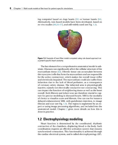
ex-vivo studies [49,55–57], and still widely used (see Fig. 1.3).
Figure 1.3. Example of heart fiber model computed using rule-based approach on
a patient-specific heart anatomy.
The last element for a comprehensive anatomical model is sub-
strate. Diseases can significantly affect the cellular structure of the
myocardium tissue [42]. Fibrotic tissue can accumulate between
the myocytes (cells that form the myocardium and are responsible
for the active contraction), which makes the overall tissue stiffer
and electrically impaired. The myocardium could also suffer from
infarction due to the lack of blood perfusion, as a consequence
of coronary artery disease. The infarcted area is physiologically
inactive, namely not electrically conductive nor contracting. This
can impact the function of neighboring tissue as well as the heart
overall. Both fibrosis and infarct scar are therefore crucial to cap-
ture for precise modeling in diseased hearts. MRI is the modality
of choice to visualize scars and fibrosis. One can use for instance
delayed-enhancement MRI, with gadolinium injection, to image
fibrosis and scar (see Fig. 1.4). The region is segmented by an ex-
pert or using image processing algorithms, and included into the
anatomical model. Chapter 2 gives more details on how this is
done in practice.
1.2 Electrophysiology modeling
Heart function is determined by the coordinated, rhythmic
contraction of the chambers, displacing blood to the body. Such
coordination requires an effective activation system that ensures
synchronized contraction. This functionality is achieved through
the cardiac electrical system, and is called electrophysiology (EP).