
Page 38 - Artificial Intelligence for Computational Modeling of the Heart
P. 38
8 Chapter 1 Multi-scale models of the heart for patient-specific simulations
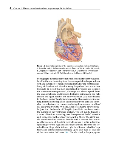
Figure 1.5. Schematic depiction of the electrical conduction system of the heart.
1. Sinoatrial node, 2. Atrioventricular node, 3. Bundle of His, 4. Left bundle branch,
5. Left posterior fascicle, 6. Left anterior fascicle, 7. Left ventricle, 8. Ventricular
septum, 9. Right ventricle, 10. Right bundle branch. (Source: Wikipedia.)
belonging to the electrical conduction system are electrically insu-
lated by fibrous sheathing from the non-specialized myocardium,
with the exception of junction points that allow the controlled de-
livery of the electrical stimulus along the path of the conduction.
It should be noted that non-specialized myocytes also conduct
the transmembrane potential, although at a slower speed. From
the sino-atrial node and through dedicated pathways in the right
atrium, the signal reaches the atrioventricular (AV) node located
in the lower part of the right atrium on the fibrous atrioventricular
ring. Fibrous tissue separates the musculature of atria and ventri-
cles, the only electrical connection being the muscular bundle of
His departing from the AV node. After crossing the atrioventricu-
lar junction, the bundle of His splits (usually in two branches) at
the summit of the ventricular septum. The left bundle branch is
a series of fascicles spreading over the septum of the left ventricle
and connecting with ordinary myocardial fibers. The right bun-
dle branch tends to remain a bundle until it reaches the anterior
papillary muscle of the right ventricle, where it splits in fascicles
spreading over the right ventricle myocardium. The tree-like ter-
minal branchings of the left and right bundles are called Purkinje
fibers and extend subendocardially up to one third or one half
of the ventricular thickness [58]. The electrical pulse propagates