
Page 34 - Artificial Intelligence for Computational Modeling of the Heart
P. 34
4 Chapter 1 Multi-scale models of the heart for patient-specific simulations
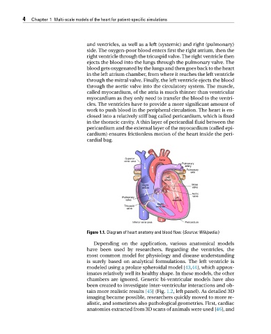
and ventricles, as well as a left (systemic) and right (pulmonary)
side. The oxygen-poor blood enters first the right atrium, then the
right ventricle through the tricuspid valve. The right ventricle then
ejects the blood into the lungs through the pulmonary valve. The
blood gets oxygenated by the lungs and then goes back to the heart
in the left atrium chamber, from where it reaches the left ventricle
through the mitral valve. Finally, the left ventricle ejects the blood
through the aortic valve into the circulatory system. The muscle,
called myocardium, of the atria is much thinner than ventricular
myocardium as they only need to transfer the blood to the ventri-
cles. The ventricles have to provide a more significant amount of
work to push blood in the peripheral circulation. The heart is en-
closed into a relatively stiff bag called pericardium, which is fixed
in the thoracic cavity. A thin layer of pericardial fluid between the
pericardium and the external layer of the myocardium (called epi-
cardium) ensures frictionless motion of the heart inside the peri-
cardial bag.
Figure 1.1. Diagram of heart anatomy and blood flow. (Source: Wikipedia.)
Depending on the application, various anatomical models
have been used by researchers. Regarding the ventricles, the
most common model for physiology and disease understanding
is surely based on analytical formulations. The left ventricle is
modeled using a prolate spheroidal model [43,44], which approx-
imates relatively well its healthy shape. In these models, the other
chambers are ignored. Generic bi-ventricular models have also
been created to investigate inter-ventricular interactions and ob-
tain more realistic results [45](Fig. 1.2, left panel). As detailed 3D
imaging became possible, researchers quickly moved to more re-
alistic, and sometimes also pathological geometries. First, cardiac
anatomies extracted from 3D scans of animals were used [46], and