
Page 109 - Biomedical Engineering and Design Handbook Volume 1, Fundamentals
P. 109
86 BIOMECHANICS OF THE HUMAN BODY
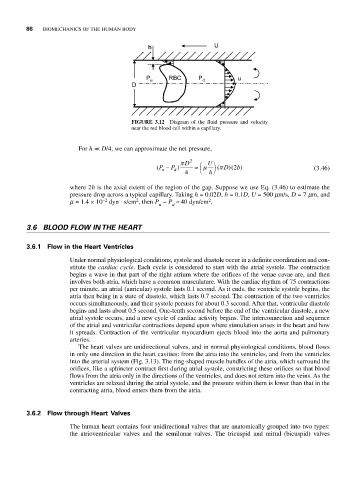
FIGURE 3.12 Diagram of the fluid pressure and velocity
near the red blood cell within a capillary.
For h << D/4, we can approximate the net pressure,
π D 2 ⎛ U ⎞
(P − P d ) ≈ μ ( D 2 ) (3.46)
π
)( b
u
4 ⎝ h ⎠
where 2b is the axial extent of the region of the gap. Suppose we use Eq. (3.46) to estimate the
pressure drop across a typical capillary. Taking h = 0.02D, b = 0.1D, U = 500 mm/s, D = 7 mm, and
.
2
2
m = 1.4 × 10 −2 dyn s/cm , then P − P ª 40 dyn/cm .
u d
3.6 BLOOD FLOW IN THE HEART
3.6.1 Flow in the Heart Ventricles
Under normal physiological conditions, systole and diastole occur in a definite coordination and con-
stitute the cardiac cycle. Each cycle is considered to start with the atrial systole. The contraction
begins a wave in that part of the right atrium where the orifices of the venae cavae are, and then
involves both atria, which have a common musculature. With the cardiac rhythm of 75 contractions
per minute, an atrial (auricular) systole lasts 0.1 second. As it ends, the ventricle systole begins, the
atria then being in a state of diastole, which lasts 0.7 second. The contraction of the two ventricles
occurs simultaneously, and their systole persists for about 0.3 second. After that, ventricular diastole
begins and lasts about 0.5 second. One-tenth second before the end of the ventricular diastole, a new
atrial systole occurs, and a new cycle of cardiac activity begins. The interconnection and sequence
of the atrial and ventricular contractions depend upon where stimulation arises in the heart and how
it spreads. Contraction of the ventricular myocardium ejects blood into the aorta and pulmonary
arteries.
The heart valves are unidirectional valves, and in normal physiological conditions, blood flows
in only one direction in the heart cavities: from the atria into the ventricles, and from the ventricles
into the arterial system (Fig. 3.13). The ring-shaped muscle bundles of the atria, which surround the
orifices, like a sphincter contract first during atrial systole, constricting these orifices so that blood
flows from the atria only in the directions of the ventricles, and does not return into the veins. As the
ventricles are relaxed during the atrial systole, and the pressure within them is lower than that in the
contracting atria, blood enters them from the atria.
3.6.2 Flow through Heart Valves
The human heart contains four unidirectional valves that are anatomically grouped into two types:
the atrioventricular valves and the semilunar valves. The tricuspid and mitral (bicuspid) valves