
Page 111 - Biomedical Engineering and Design Handbook Volume 1, Fundamentals
P. 111
88 BIOMECHANICS OF THE HUMAN BODY
1994). More recently, fluid-structure interaction models, based on the immersed boundary technique,
were used for describing valvular function (Sauob et al., 1999). In these models, strong coupled
fluid-structure dynamic simulations were obtained. These models allowed for the inclusion of bend-
ing stresses, contact between adjacent leaflets when they coapted, and transient three-dimensional
blood flow through the valve.
3.6.3 Coronary Blood Flow
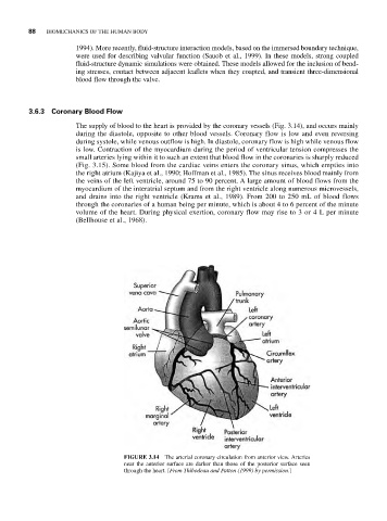
The supply of blood to the heart is provided by the coronary vessels (Fig. 3.14), and occurs mainly
during the diastole, opposite to other blood vessels. Coronary flow is low and even reversing
during systole, while venous outflow is high. In diastole, coronary flow is high while venous flow
is low. Contraction of the myocardium during the period of ventricular tension compresses the
small arteries lying within it to such an extent that blood flow in the coronaries is sharply reduced
(Fig. 3.15). Some blood from the cardiac veins enters the coronary sinus, which empties into
the right atrium (Kajiya et al., 1990; Hoffman et al., 1985). The sinus receives blood mainly from
the veins of the left ventricle, around 75 to 90 percent. A large amount of blood flows from the
myocardium of the interatrial septum and from the right ventricle along numerous microvessels,
and drains into the right ventricle (Krams et al., 1989). From 200 to 250 mL of blood flows
through the coronaries of a human being per minute, which is about 4 to 6 percent of the minute
volume of the heart. During physical exertion, coronary flow may rise to 3 or 4 L per minute
(Bellhouse et al., 1968).
FIGURE 3.14 The arterial coronary circulation from anterior view. Arteries
near the anterior surface are darker than those of the posterior surface seen
through the heart. [From Thibodeau and Patton (1999) by permission.]