
Page 182 - Biomedical Engineering and Design Handbook Volume 1, Fundamentals
P. 182
BIOMECHANICS OF THE MUSCULOSKELETAL SYSTEM 159
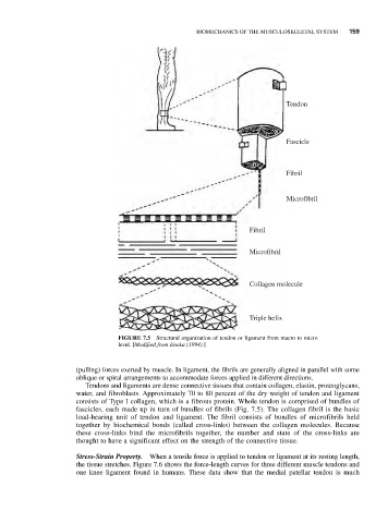
Tendon
Fascicle
Fibril
Microfibril
Fibril
Microfibril
Collagen molecule
Triple helix
FIGURE 7.5 Structural organization of tendon or ligament from macro to micro
level. [Modified from Enoka (1994).]
(pulling) forces exerted by muscle. In ligament, the fibrils are generally aligned in parallel with some
oblique or spiral arrangements to accommodate forces applied in different directions.
Tendons and ligaments are dense connective tissues that contain collagen, elastin, proteoglycans,
water, and fibroblasts. Approximately 70 to 80 percent of the dry weight of tendon and ligament
consists of Type I collagen, which is a fibrous protein. Whole tendon is comprised of bundles of
fascicles, each made up in turn of bundles of fibrils (Fig. 7.5). The collagen fibril is the basic
load-bearing unit of tendon and ligament. The fibril consists of bundles of microfibrils held
together by biochemical bonds (called cross-links) between the collagen molecules. Because
these cross-links bind the microfibrils together, the number and state of the cross-links are
thought to have a significant effect on the strength of the connective tissue.
Stress-Strain Property. When a tensile force is applied to tendon or ligament at its resting length,
the tissue stretches. Figure 7.6 shows the force-length curves for three different muscle tendons and
one knee ligament found in humans. These data show that the medial patellar tendon is much