
Page 326 - Biomedical Engineering and Design Handbook Volume 1, Fundamentals
P. 326
ELECTROMYOGRAPHY AS A TOOL TO ESTIMATE MUSCLE FORCES 303
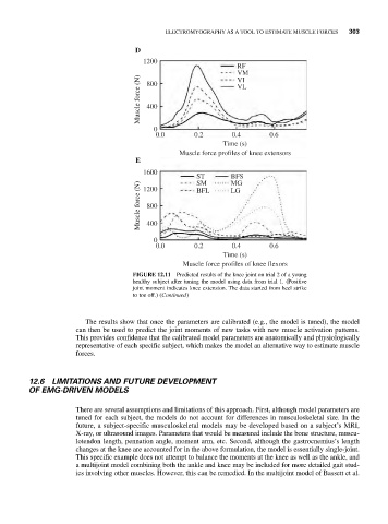
D
1200
RF
VM
Muscle force (N) 400 VL
VI
800
0
0.0 0.2 0.4 0.6
Time (s)
Muscle force profiles of knee extensors
E
1600
ST BFS
MG
SM
Muscle force (N) 800 BFL LG
1200
400
0
0.0 0.2 0.4 0.6
Time (s)
Muscle force profiles of knee flexors
FIGURE 12.11 Predicted results of the knee joint on trial 2 of a young
healthy subject after tuning the model using data from trial 1. (Positive
joint moment indicates knee extension. The data started from heel strike
to toe off.) (Continued)
The results show that once the parameters are calibrated (e.g., the model is tuned), the model
can then be used to predict the joint moments of new tasks with new muscle activation patterns.
This provides confidence that the calibrated model parameters are anatomically and physiologically
representative of each specific subject, which makes the model an alternative way to estimate muscle
forces.
12.6 LIMITATIONS AND FUTURE DEVELOPMENT
OF EMG-DRIVEN MODELS
There are several assumptions and limitations of this approach. First, although model parameters are
tuned for each subject, the models do not account for differences in musculoskeletal size. In the
future, a subject-specific musculoskeletal models may be developed based on a subject’s MRI,
X-ray, or ultrasound images. Parameters that would be measured include the bone structure, muscu-
lotendon length, pennation angle, moment arm, etc. Second, although the gastrocnemius’s length
changes at the knee are accounted for in the above formulation, the model is essentially single-joint.
This specific example does not attempt to balance the moments at the knee as well as the ankle, and
a multijoint model combining both the ankle and knee may be included for more detailed gait stud-
ies involving other muscles. However, this can be remedied. In the multijoint model of Bassett et al.