
Page 265 - Biomedical Engineering and Design Handbook Volume 2, Applications
P. 265
DESIGN OF MAGNETIC RESONANCE SYSTEMS 243
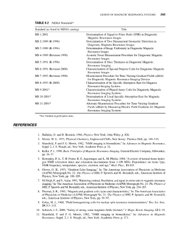
TABLE 8.2 NEMA Standards*
Standard (as listed in NEMA catalog) Title
MS 1-2001 Determination of Signal-to-Noise Ratio (SNR) in Diagnostic
Magnetic Resonance Images
MS 2-1989 (R-1996) Determination of Two-Dimensional Geometric Distortion in
Diagnostic Magnetic Resonance Images
MS 3-1989 (R-1994) Determination of Image Uniformity in Diagnostic Magnetic
Resonance Images
MS 4-1989 (Revision 1998) Acoustic Noise Measurement Procedure for Diagnostic Magnetic
Resonance Images
MS 5-1991 (R-1996) Determination of Slice Thickness in Diagnostic Magnetic
Resonance Imaging
MS 6-1991 (Revision 2000) Characterization of Special Purpose Coils for Diagnostic Magnetic
Resonance Images
MS 7-1993 (Revision 1998) Measurement Procedure for Time-Varying Gradient Fields (dB/dt)
for Diagnostic Magnetic Resonance Imaging Devices
MS 8-1993 (R-2000) Characterization of the Specific Absorption Rate for Magnetic
Resonance Imaging Systems
MS 9-2001* Characterization of Phased Array Coils for Diagnostic Magnetic
Resonance Imaging Systems
MS 10-2001* Determination of Local Specific Absorption Rate for Magnetic
Resonance Imaging Systems
MS 11-2001* Alternate Measurement Procedure for Time-Varying Gradient
Fields (dB/dt) by Measuring Electric Field Gradients for Magnetic
Resonance Imaging Systems
*Not finished at publication time.
REFERENCES
1. Halliday, D. and R. Resnick, 1966, Physics, New York: John Wiley, p. 826.
2. Moore, W. J., 1972, Physical Chemistry, Englewood Cliffs, New Jersey: Prentice Hall, pp. 140–143.
3. Mansfield, P. and P. G. Morris, 1982, “NMR imaging in biomedicine,” In: Advances in Magnetic Resonance,
Suppl. 2, J. S. Waugh, ed., New York: Academic Press, p. 32.
4. Keller, P. J., 1990, Basic Principles of Magnetic Resonance Imaging, General Electric Company, Milwaukee,
pp. 16–37.
5. Bottomley, P. A., T. H. Foster, R. E. Argersinger, and L. M. Pfiefer, 1984, “A review of normal tissue hydro-
gen NMR relaxation times and relaxation mechanisms from 1-100 MHz: Dependence on tissue type,
NMR frequency, temperature, species, excision, and age,” Med. Phys., 11:425.
6. Glover, G. H., 1993, “Gradient Echo Imaging,” In: The American Association of Physicists in Medicine
(AAPM) Monograph No. 21: The Physics of MRI, P. Sprawls and M. Bronskill, eds., American Institute of
Physics, New York, pp. 188–205.
7. McVeigh, E. and E. Atalar, 1993, “Balancing contrast, Resolution, and signal-to-noise ratio in magnetic resonance
imaging,” In: The American Association of Physicists in Medicine (AAPM) Monograph No. 21: The Physics of
MRI, P. Sprawls and M. Bronskill, eds., American Institute of Physics, New York, pp. 234–267.
8. Thomas, S. R., 1993, “Magnets and gradient coils: types and characteristics,” In: The American Association
of Physicists in Medicine (AAPM) Monograph No. 21: The Physics of MRI, P. Sprawls and M. Bronskill,
eds., American Institute of Physics, New York, pp. 56–97.
9. Golay, M. J., 1968, “Field homogenizing coils for nuclear spin resonance instrumentation,” Rev. Sci. Inst.,
29:313–315.
10. Schenck, J. F., 2000, “Safety of strong, static magnetic fields (invited),” J. Magn. Reson. Imaging, 12:2–19.
11. Mansfield, P. and P. G. Morris, 1982, “NMR imaging in biomedicine,” In: Advances in Magnetic
Resonance, Suppl. 2, J. S. Waugh, ed., New York: Academic Press, p. 271.