
Page 332 - Biomedical Engineering and Design Handbook Volume 2, Applications
P. 332
310 DIAGNOSTIC EQUIPMENT DESIGN
10.4.5 Ancillary Instrumentation
Data Acquisition. The broad interpretation of data acquisition relates to the various operations that
are executed in a sequential fashion to secure a complete data set for subsequent computational
reconstruction. In a narrower sense, this principally involves specimen motion, frame grabbing, and
storing. For an efficient operation, these functions must be carried out automatically with provision
for programming operational parameters such as the motion step value and total number of
steps/frames, and the camera integration period. The system described in this section has an NT PC
with an installed programmable PCI board and a fiber-optic link to control the camera and also com-
municates with a programmable servo controller for specimen motion via the RS232 port.
The organization of frame grabbing and specimen motion is performed by a programmable inter-
face written in Python, which is an interpreted, interactive, object-oriented language. This provides
the handshaking function that is essential for error-free sequential operation. The interface will pick
up configuration files that set the camera parameters and servo control parameters. Run-specific
details are entered in proffered dialog boxes at the outset of a data acquisition sequence. The image
frames are temporarily stored on the PC hard disk and subsequently downloaded onto a Sun Micro
Systems SCSI multiple-disk drive.
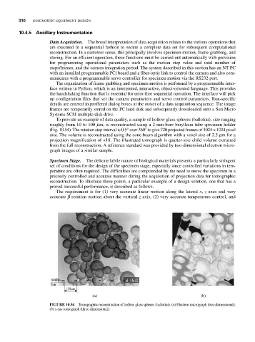
To provide an example of data quality, a sample of hollow glass spheres (ballotini), size ranging
roughly from 10 to 100 mm, is reconstructed using a 2-mm-bore beryllium tube specimen holder
(Fig. 10.54). The rotation step interval is 0.5° over 360° to give 720 projected frames of 1024 × 1024 pixel
size. The volume is reconstructed using the cone-beam algorithm with a voxel size of 2.5 mm for a
projection magnification of ×18. The illustrated tomograph is quarter-size child volume extracted
from the full reconstruction. A reference standard was provided by two-dimensional electron micro-
graph images of a similar sample.
Specimen Stage. The delicate labile nature of biological materials presents a particularly stringent
set of conditions for the design of the specimen stage, especially since controlled variations in tem-
perature are often required. The difficulties are compounded by the need to move the specimen in a
precisely controlled and accurate manner during the acquisition of projection data for tomographic
reconstruction. To illustrate these points, a particular example of a design solution, one that has a
proved successful performance, is described as follows.
The requirement is for (1) very accurate linear motion along the lateral x, z axes and very
accurate b rotation motion about the vertical z axis, (2) very accurate temperature control, and
FIGURE 10.54 Tomographic reconstruction of hollow glass spheres (ballotini). (a) Electron micrograph (two-dimensional);
(b) x-ray tomograph (three-dimensional).