
Page 327 - Biomedical Engineering and Design Handbook Volume 2, Applications
P. 327
THE PRINCIPLES OF X-RAY COMPUTED TOMOGRAPHY 305
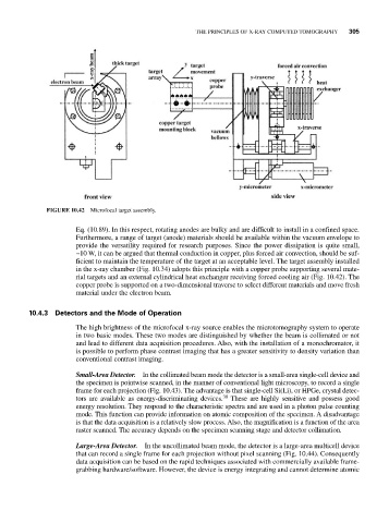
FIGURE 10.42 Microfocal target assembly.
Eq. (10.89). In this respect, rotating anodes are bulky and are difficult to install in a confined space.
Furthermore, a range of target (anode) materials should be available within the vacuum envelope to
provide the versatility required for research purposes. Since the power dissipation is quite small,
~10 W, it can be argued that thermal conduction in copper, plus forced air convection, should be suf-
ficient to maintain the temperature of the target at an acceptable level. The target assembly installed
in the x-ray chamber (Fig. 10.34) adopts this principle with a copper probe supporting several mate-
rial targets and an external cylindrical heat exchanger receiving forced cooling air (Fig. 10.42). The
copper probe is supported on a two-dimensional traverse to select different materials and move fresh
material under the electron beam.
10.4.3 Detectors and the Mode of Operation
The high brightness of the microfocal x-ray source enables the microtomography system to operate
in two basic modes. These two modes are distinguished by whether the beam is collimated or not
and lead to different data acquisition procedures. Also, with the installation of a monochromator, it
is possible to perform phase contrast imaging that has a greater sensitivity to density variation than
conventional contrast imaging.
Small-Area Detector. In the collimated beam mode the detector is a small-area single-cell device and
the specimen is pointwise scanned, in the manner of conventional light microscopy, to record a single
frame for each projection (Fig. 10.43). The advantage is that single-cell Si(Li), or HPGe, crystal detec-
tors are available as energy-discriminating devices. 30 These are highly sensitive and possess good
energy resolution. They respond to the characteristic spectra and are used in a photon pulse counting
mode. This function can provide information on atomic composition of the specimen. A disadvantage
is that the data acquisition is a relatively slow process. Also, the magnification is a function of the area
raster scanned. The accuracy depends on the specimen scanning stage and detector collimation.
Large-Area Detector. In the uncollimated beam mode, the detector is a large-area multicell device
that can record a single frame for each projection without pixel scanning (Fig. 10.44). Consequently
data acquisition can be based on the rapid techniques associated with commercially available frame-
grabbing hardware/software. However, the device is energy integrating and cannot determine atomic