Page 324 - Biomedical Engineering and Design Handbook Volume 2, Applications
P. 324

302  DIAGNOSTIC EQUIPMENT DESIGN

                       This is a very inefficient process with most of the energy appearing as heat. As a consequence, the
                       temperature of the target material can reach melting point. The liquid metal is subsequently removed
                       by electron beam pressure, resulting in the excavation of a pit or groove. This degradation of the tar-
                       get material creates an enlarged focal spot. Therefore, it is important to determine the maximum
                       power density that a target material can safely accommodate in order to produce maximum x-ray
                       flux to satisfy the threshold of detection condition [Eq. (10.91)].
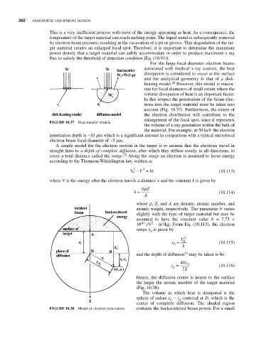
                                                              For the large-focal-diameter electron beams
                                                           associated with medical x-ray sources, the heat
                                                           dissipation is considered to occur at the surface
                                                           and the analytical geometry is that of a disk-
                                                           heating model. 28  However, this model is inaccu-
                                                           rate for focal diameters of small extent where the
                                                           volume dissipation of heat is an important factor.
                                                           In this respect the penetration of the beam elec-
                                                           trons into the target material must be taken into
                                                           account (Fig. 10.37). Furthermore, the extent of
                                                           the electron distribution will contribute to the
                                                           enlargement of the focal spot, since it represents
                       FIGURE 10.37  Heat transfer models.
                                                           the volume of x-ray generation within the bulk of
                                                           the material. For example, at 50 keV the electron
                       penetration depth is ~10 mm which is a significant amount in comparison with a typical microfocal
                       electron beam focal diameter of ~5 mm.
                         A simple model for the electron motion in the target is to assume that the electrons travel in
                       straight lines to a depth of complete diffusion, after which they diffuse evenly in all directions, to
                                                   29
                       cover a total distance called the range. Along the range an electron is assumed to loose energy
                       according to the Thomson-Whiddington law, written as
                                                           2
                                                       2
                                                      V − V =  kx                       (10.113)
                                                       0
                       where V is the energy after the electron travels a distance x and the constant k is given by
                                                           ρ
                                                          bZ
                                                       k =                              (10.114)
                                                           A
                                                        where r, Z, and A are density, atomic number, and
                                                        atomic weight, respectively. The parameter b varies
                                                        slightly with the type of target material but may be
                                                        assumed to have the constant value  b = 7.75  ×
                                                                  2
                                                              2
                                                        10 10  eV ⋅ m /kg. From Eq. (10.113), the electron
                                                        range x is given by
                                                              0
                                                                          V  2
                                                                      x =  0            (10.115)
                                                                       0
                                                                          k
                                                                           29
                                                        and the depth of diffusion may be taken to be
                                                                         40 x
                                                                      z =   0           (10.116)
                                                                       d
                                                                          7 Z
                                                        Hence, the diffusion center is nearer to the surface
                                                        the larger the atomic number of the target material
                                                        (Fig. 10.38).
                                                           The volume in which heat is dissipated is the
                                                        sphere of radius x − z centered at D, which is the
                                                                      0
                                                                         d
                                                        center of complete diffusion.  The shaded region
                       FIGURE 10.38  Model of electron penetration.  contains the backscattered beam power. For a small
   319   320   321   322   323   324   325   326   327   328   329