
Page 90 - Biomedical Engineering and Design Handbook Volume 2, Applications
P. 90
OVERVIEW OF CARDIOVASCULAR DEVICES 69
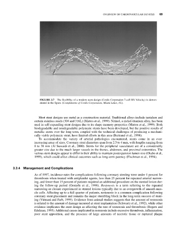
FIGURE 3.7 The flexibility of a modern stent design (Cordis Corporation 7-cell BX Velocity) is demon-
strated in the figure. (Compliments of Cordis Corporation, Miami Lakes, FL).
Most stent designs use metal as a construction material. Traditional alloys include tantalum and
certain stainless steels (304 and 316L) (Mattos et al., 1999). Nitinol, a nickel-titanium alloy, has been
used in self-expanding stent designs due to its shape memory properties (Mattos et al., 1999). Both
biodegradable and nondegradable polymeric stents have been developed, but the positive results of
metallic stents over the long term, coupled with the technical challenges of producing a mechani-
cally viable polymeric stent, have limited efforts in this area (Bertrand et al., 1998).
To accommodate the variety of arterial pathologies encountered, stents come in an ever-
increasing array of sizes. Coronary stent diameters span from 2.5 to 4 mm, with lengths ranging from
8 to 38 mm (Al Suwaidi et al., 2000). Stents for the peripheral vasculature are of a considerably
greater size due to the much larger vessels in the thorax, abdomen, and proximal extremities. The
various stent designs appear to differ in their ability to maintain postexpansion lumen size (Okabe et al.,
1999), which could affect clinical outcomes such as long-term patency (Fischman et al., 1994).
3.3.4 Management and Complications
As of 1997, incidence rates for complications following coronary stenting were under 1 percent for
thrombosis when treated with antiplatelet agents, less than 25 percent for repeated arterial narrow-
ing, and fewer than 15 percent of patients required an additional procedure on the stented lesion dur-
ing the follow-up period (Oesterle et al., 1998). Restenosis is a term referring to the repeated
narrowing or closure experienced in stented lesions typically due to an overgrowth of smooth mus-
cle cells. Affecting up to a full quarter of patients, restenosis is a common complication following
coronary stent placement and remains the major stumbling block in the long-term success of stent-
ing (Virmani and Farb, 1999). Evidence from animal studies suggests that the amount of restenosis
is related to the amount of damage incurred at stent implantation (Schwartz et al., 1992), while other
evidence implicates the stent design as affecting the rate of restenosis and thrombosis (Rogers and
Edelman, 1995). Additional causes implicated in restenosis include excessive thrombosis, inflammation,
poor stent apposition, and the presence of large amounts of necrotic tissue or ruptured plaque