
Page 167 - Carbon Nanotubes
P. 167
158 Y. SAITO
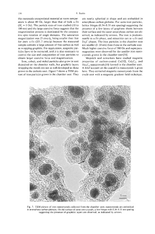
this nanoscale encapsulated material at room temper- are nearly spherical in shape and are embedded in
ature is about 80 Oe, larger than that of bulk a-Fe amorphous carbon globules. For some iron particles,
(H, = 1 Oe). The particle sizes of iron studied (10 to lattice fringes (0.34-0.35 nm spacing) suggesting the
100 nm) and the large coercive force suggests that the presence of a few layers of graphene sheets between
magnetization process is dominated by the coopera- their surface and the outer amorphous carbon are ob-
tive spin rotation of single domains. The saturation served, as indicated by arrows. The iron is predomi-
magnetization was 25 emu/g, being smaller than that nantly in a-Fe phase, and minorities are in y-Fe and
for pure a-Fe (221.7 emu/g) because the measured Fe3C phases. The iron particles in the chamber soot
sample contains a large amount of free carbon as well are smaller (3-10 nm) than those in the cathode soot.
as wrapping graphite. For application, magnetic par- Much higher coercive force of 380 Oe and superpara-
ticles have to be extracted, and it is also necessary to magnetism were observed for the smaller iron nano-
control the size and composition of iron particles to crystals grown in the chamber soot[31].
obtain larger coercive force and magnetization. Majetich and coworkers have studied magnetic
Iron, cobalt, and nickel particles also grow in soot properties of carbon-coated Co[32], Gd2C3, and
deposited on the chamber walls, but graphitic layers Ho& nanocrystals[33] formed in the chamber soot.
wrapping the metals are not so well-developed as those A brief account on the coated Co nanocrystals is given
grown in the cathode soot. Figure 7 shows a TEM pic- here. They extracted magnetic nanocrystals from the
ture of iron particles grown in the chamber soot. They crude soot with a magnetic gradient field technique.
Fig. 7. TEM picture of iron nanocrystals collected from the chamber soot; nanocrystals are embedded
in amorphous carbon globules. On the surface of some core crystals, a few fringes with 0.34-0.35 nm spacing
suggesting the presence of graphitic layers are observed, as indicated by arrows.