
Page 168 - Carbon Nanotubes
P. 168
Nanoparticles and filled nanocapsules 159
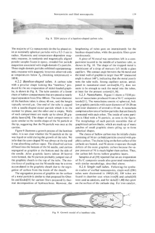
Fig. 8. TEM picture of a bamboo-shaped carbon tube.
The majority of Co nanocrystals (in the fcc phase) ex- lengthening of tubes goes on intermittently for the
ist as nominally spherical particles with a 0.5-5 nm in bamboo-shaped tubes, while the pyrolytic fibers grow
radius. Hysteretic and temperature-dependent mag- continuously.
netic response, in randomly and magnetically aligned A piece of Ni metal was sometimes left in a com-
powder samples frozen in epoxy, revealed fine particle partment located in the middle of a bamboo tube, as
magnetism associated with single-domain Co particles. shown in Fig. 10. The shape of the trapped metal is
These single-domain particles exhibited superparamag- reminiscent of a drop of mercury left inside a glass
netic response with magnetic hysteresis observed only capillary. The contact angle between the Ni metal and
at temperatures below Ts (blocking temperature) = the inner wall of graphite is larger than 90” (measured
160 K. angle is about 140”), indicating that the metal poorly
4.2.2 Bamboo-shaped tubes. A carbon tube wets the tube walls. Strong capillary action, antici-
with a peculiar shape looking like “bamboo,” pro- pated in nanometer-sized cavities[36,37], does not
duced by the arc evaporation of nickel-loaded graph- seem to be enough to suck the metal into the tubes,
ite, is shown in Fig. 8. The tube consists of a linear at least for the present system[ll,38].
chain of hollow compartments that are spaced at nearly 4.2.3 Nanochains. Figure 11 shows a TEM pic-
equal separation from 50 to 100 nm. The outer diameter ture of nanochains produced from a Ni/C composite
of the bamboo tubes is about 40 nm, and the length anode[ll]. The nanochains consist of spherical, hol-
typically several pm. One end of the tube is capped low graphitic particles with outer diameters of 10-20 nm
with a needle-shaped nickel particle which is in the and inner diameters of several to 10 nm. A nanochain
normal fcc phase, and the other end is empty. Walls comprises a few tens of hollow particles that are linearly
of each compartment are made up by about 20 gra- connected with each other. The inside of some parti-
phitic layers[34]. The shape of each compartment is cles is filled with a Ni particle, as seen in the figure.
quite similar to the needle-shape of the Ni particle at The morphology of each particle resembles that of
the tip, suggesting that the Ni particle was once at the graphitized carbon blacks, which are made up of many
cavities. patches of small graphitic sheets piling up to form
Figure 9 illustrates a growth process of the bamboo spherical shapes.
tubes. It is not clear whether the Ni particle at the tip The chains of hollow carbon may be initially chains
was liquid or solid during the growth of the tube. We consisting of Ni (or carbide) particles covered with gra-
infer that the cone-shaped Ni was always at the tip and phitic carbon. The chains lying on the hot surface of the
it was absorbing carbon vapor. The dissolved carbon cathode are heated, and Ni atoms evaporate through
diffused into the bottom of the Ni needle, and carbon defects of the outer graphitic carbon because the va-
segregated as graphite at the bottom and the side of por pressure of Ni is much higher than carbon. Thus,
the needle. After graphitic layers (about 20 layers) the carbon left forms hollow graphitic layers.
were formed, the Ni particle probably jumped out of Seraphin el al.[39] reported that an arc evaporation
the graphitic sheath to the top of the tube. The mo- of Fe/C composite anode also generated nanochains
tive force of pushing out the Ni needle may be a stress with similar morphology, described above.
accumulated in the graphitic sheath due to the segre- 4.2.4 Single-wall tubes. Following the synthe-
gation of carbon from the inside of the sheath. sis studies of stuffed nanocapsules, single-wall (SW)
The segregation process of graphite on the surface tubes were discovered in 1993[9,10]. SW tubes are
of a metal particle is similar to that proposed by Ober- found in chamber soot when iron[9] and cobalt[lO]
lin and Endo[35] for carbon fibers prepared by ther- were used as catalysts, and for nickel[ 11,401 they grow
mal decomposition of hydrocarbons. However, the on the surface of the cathode slag. For iron catalyst,
Fig. 9. Growth model of a bamboo tube.