
Page 173 - Carbon Nanotubes
P. 173
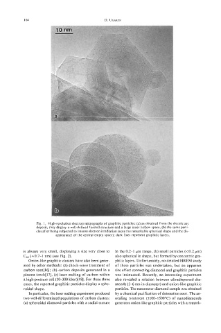
164                                   D. UGARTE


                         i


                  Fig.  1.  High-resolution  electron micrographs of graphitic particles: (a) as obtained from the electric arc
                  deposit, they display a well-defined faceted structure and a large inner hollow space, (b) the same parti-
                  cles after being subjected to intense electron irradiation (note the remarkable spherical shape and the dis-
                           appearance of the central empty space); dark lines represent graphitic layers.


            is always very small, displaying a size very close to   in the 0.2-1  pm range, (b) small particles (<0.2 pm)
            C60 (20.7-1  nm) (see Fig. 2).             also spherical in shape, but formed by concentric gra-
               Onion-like graphitic clusters have also been gener-   phitic layers. Unfortunately, no detailed HREM study
            ated by other methods: (a) shock-wave treatment  of   of  these particles was undertaken, but  an apparent
            carbon soot[l6]; (b) carbon deposits generated in a   size effect connecting diamond and graphitic particles
            plasma torch[l7], (c) laser melting of carbon within   was insinuated.  Recently,  an interesting  experiment
            a high-pressure cell (50-300  kbar)[l8]. For these three   also revealed a  relation  between ultradispersed dia-
            cases, the reported graphitic particles display a sphe-   monds (3-6 nm in diameter) and onion-like graphitic
            roidal shape.                              particles. The nanometer diamond sample was obtained
               In particular, the laser melting experiment produced   by a chemical purification of detonation soot. The an-
            two well-differentiated populations of carbon clusters:  nealing  treatment (1 100-1 500°C) of  nanodiamonds
            (a) spheroidal diamond particles with a radial texture   generates onion-like graphitic particles with a remark-