Page 158 - Computational Modeling in Biomedical Engineering and Medical Physics
P. 158
Bioimpedance methods 147
membranes, proteins, and other organic macromolecules are produced, and (iii) in the
range of gigahertz (called “γ dispersion region”) the polarization of water molecules hap-
pens; both β and γ dispersion regions are characterized by orientational polarization.
To cope with this wide spectrum of frequency-related processes, several macro-
scopic models of permittivity were proposed. For example, the single-dispersion
Cole Cole relaxation model (Cole and Cole, 1941) presents the complex relative per-
mittivity as frequency dependent function for three tissues of the body (skin, muscle,
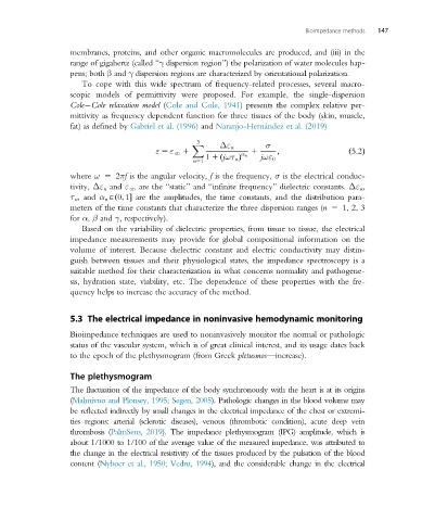
fat) as defined by Gabriel et al. (1996) and Naranjo-Hernández et al. (2019)
3
X Δε n σ
ε 5 ε N 1 1 ; ð5:2Þ
α n
1 1 jωτ n Þ jωε 0
ð
n51
where ω 5 2πf is the angular velocity, f is the frequency, σ is the electrical conduc-
tivity, Δε n and ε N are the “static” and “infinite frequency” dielectric constants. Δε n ,
τ n , and α n A 0; 1ð are the amplitudes, the time constants, and the distribution para-
meters of the time constants that characterize the three dispersion ranges (n 5 1, 2, 3
for α, β and γ, respectively).
Based on the variability of dielectric properties, from tissue to tissue, the electrical
impedance measurements may provide for global compositional information on the
volume of interest. Because dielectric constant and electric conductivity may distin-
guish between tissues and their physiological states, the impedance spectroscopy is a
suitable method for their characterization in what concerns normality and pathogene-
sis, hydration state, viability, etc. The dependence of these properties with the fre-
quency helps to increase the accuracy of the method.
5.3 The electrical impedance in noninvasive hemodynamic monitoring
Bioimpedance techniques are used to noninvasively monitor the normal or pathologic
status of the vascular system, which is of great clinical interest, and its usage dates back
to the epoch of the plethysmogram (from Greek pletusmos—increase).
The plethysmogram
The fluctuation of the impedance of the body synchronously with the heart is at its origins
(Malmivuo and Plonsey, 1995; Segen, 2005). Pathologic changes in the blood volume may
be reflected indirectly by small changes in the electrical impedance of the chest or extremi-
ties regions: arterial (sclerotic diseases), venous (thrombotic condition), acute deep vein
thrombosis (PalmSens, 2019). The impedance plethysmogram (IPG) amplitude, which is
about 1/1000 to 1/100 of the average value of the measured impedance, was attributed to
thechangeinthe electrical resistivityofthe tissues produced by the pulsation of the blood
content (Nyboer et al., 1950; Vedru, 1994), and the considerable change in the electrical

