Page 162 - Computational Modeling in Biomedical Engineering and Medical Physics
P. 162
Bioimpedance methods  151


                   2008; Osypka, 2009, Grollmuss et al., 2012; Henry et al., 2012)iseducedfromgeneral car-
                   diac output observances and injury espial of myocardial ischemic (Mellert et al., 2011).
                   EVM model analyses the change in impedance of the aortic blood soon after aortic valve
                   opening, due to the red blood cells (erythrocytes) alignment with the flow.
                      The observed EVM/ECM bioimpedance signal bears also the concurring effects of
                   respiration and fluctuations in the cardiac cycle: Z(t) 5 Z 0 1 ΔZ R 1 ΔZ C ,where Z 0
                   is the “quasi-static” part, referred to as the base impedance. ΔZ R accounts for the effects
                   of respiration and it is suppressed to the estimate SV. ΔZ C is caused by changes due to
                   the cardiac cycle, produced by the thoracic fluids, the thoracic blood volume included.
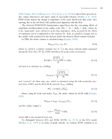
                      In TEB, the stroke volume is calculated using (Osypka, 2009)

                                               SV TEB 5 C p Uv FT UFT;                    ð5:9Þ
                                                          21
                   where C p [mL] is a patient constant, v FT [s  ] is the mean velocity index measured
                   during the flow time, FT [s]. EVM introduces the peak aortic acceleration


                                                      dZ tðÞ

                                                       dt
                                           ICON 5          min  3 1000;                  ð5:10Þ
                                                        Z 0
                   and uses it to calculate v FT yielding
                                                       v ffiffiffiffiffiffiffiffiffiffiffiffiffiffiffiffiffiffiffiffiffiffi

                                                       u
                                                            dZ tðÞ
                                                       u
                                                            dt
                                                       t
                                              v FT;EVM 5        min  ;                   ð5:11Þ
                                                             Z 0
                   and “corrects” the flow time rate, which is calculated using the left-ventricular ejec-
                   tion time, LVET and the ECG R-R interval, T RR , yielding
                                                            p ffiffiffiffiffiffiffiffiffi
                                               FT C 5 LVET= T RR ;                       ð5:12Þ
                      Hence, using the body mass index, V EPT , the stroke volume by ECM yields (Osypka,
                   2009)

                                            SV EVM 5 V EPT Uv FT;EVM UFT C ;             ð5:13Þ

                   and the cardiac output is

                                                     SV EVM
                                               CO 5         3 HR;                        ð5:14Þ
                                                      1000
                   where HR is the measured heart rate.
                      To distinguish between ICG and EVM, ICON, Eq. (5.10), in the ICG model
                   (Woltjer et al., 1997) is an index of peak velocity. In contrast, EVM considers it an

