
Page 164 - Computational Modeling in Biomedical Engineering and Medical Physics
P. 164
Bioimpedance methods 153
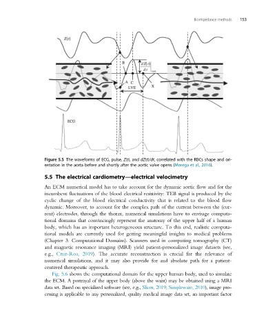
Figure 5.5 The waveforms of ECG, pulse, Z(t), and dZ(t)/dt, correlated with the RBCs shape and ori-
entation in the aorta before and shortly after the aortic valve opens (Morega et al., 2016).
5.5 The electrical cardiometry—electrical velocimetry
An ECM numerical model has to take account for the dynamic aortic flow and for the
incumbent fluctuations of the blood electrical resistivity: TEB signal is produced by the
cyclic change of the blood electrical conductivity that is related to the blood flow
dynamic. Moreover, to account for the complex path of the current between the (cur-
rent) electrodes, through the thorax, numerical simulations have to envisage computa-
tional domains that convincingly represent the anatomy of the upper half of a human
body, which has an important heterogeneous structure. To this end, realistic computa-
tional models are currently used for getting meaningful insights to medical problems
(Chapter 3: Computational Domains). Scanners used in computing tomography (CT)
and magnetic resonance imaging (MRI) yield patient-personalized image datasets (see,
e.g., Cruz-Roa, 2019). The accurate reconstruction is crucial for the relevance of
numerical simulations, and it may also provide for and absolute path for a patient-
centered therapeutic approach.
Fig. 5.6 shows the computational domain for the upper human body, used to simulate
theECM.Aportrayalofthe upperbody(abovethe waist) maybe obtainedusing aMRI
data set. Based on specialized software (see, e.g., Slicer, 2019; Simpleware, 2010), image pro-
cessing is applicable to any personalized, quality medical image data set, an important factor