
Page 96 - Computational Modeling in Biomedical Engineering and Medical Physics
P. 96
84 Computational Modeling in Biomedical Engineering and Medical Physics
(A) (B) (C)
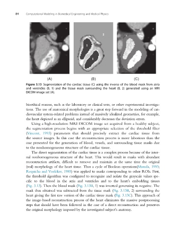
Figure 3.13 Segmentation of the cardiac tissue (C) using the inverse of the blood mask from atria
and ventricles (B, 1) and the tissue mask surrounding the heart (B, 2) generated using an MRI
DICOM image set (A).
bioethical reasons, such as the laboratory or clinical tests, or other experimental investiga-
tions. The use of anatomical morphologies is a great step forward in the modeling of car-
diovascular system-related problems instead of massively idealized geometries, for example,
the heart depicted as an ellipsoid, and considerably decreases the deviation errors.
Using a high-resolution MRI DICOM image set acquired from a healthy subject,
the segmentation process begins with an appropriate selection of the threshold filter
(Vincent, 1993) parameters that should precisely extract the cardiac tissue from
the source images. In this case the reconstruction process is more laborious than the
one presented for the generation of blood, vessels, and surrounding tissue masks due
to the nonhomogeneous structure of the cardiac tissue.
The direct segmentation of the cardiac tissue is a complex process because of the inter-
nal nonhomogeneous structure of the heart. This would result in masks with abundant
reconstruction artifacts, difficult to remove and maintain at the same time the original
(real) morphology of the heart tissue. Thus a cycle of Boolean operations(Masuda, 1993;
Requicha and Voelcker, 1985) was applied to masks corresponding to other ROIs. First,
the threshold algorithm was configured to recognize and isolate the grayscale values spe-
cific to the blood in the atria and ventricles and to the heart’s embedding tissues
(Fig. 3.13). Then the blood mask (Fig. 3.13B, 1) was inverted generating its negative. The
mask thus obtained was subtracted from the tissue mask (Fig. 3.13B, 2) surrounding the
heart giving the first raw version of the cardiac tissue mask (Fig. 3.13C). This approach of
the image-based reconstruction process of the heart eliminates the massive postprocessing
steps that should have been followed in the case of a direct reconstruction and preserves
the original morphology imposed by the investigated subject’sanatomy.