
Page 92 - Computational Modeling in Biomedical Engineering and Medical Physics
P. 92
80 Computational Modeling in Biomedical Engineering and Medical Physics
(A) (B)
(C) (D)
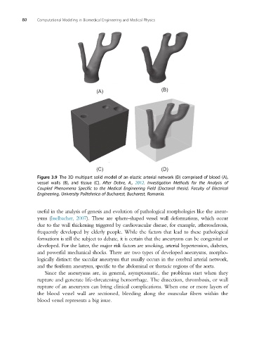
Figure 3.9 The 3D multipart solid model of an elastic arterial network (D) comprised of blood (A),
vessel walls (B), and tissue (C). After Dobre, A., 2012. Investigation Methods for the Analysis of
Coupled Phenomena Specific to the Medical Engineering Field (Doctoral thesis). Faculty of Electrical
Engineering, University Politehnica of Bucharest, Bucharest, Romania.
useful in the analysis of genesis and evolution of pathological morphologies like the aneur-
ysms (Isselbacher, 2007). These are sphere-shaped vessel wall deformations, which occur
due to the wall thickening triggered by cardiovascular disease, for example, atherosclerosis,
frequently developed by elderly people. While the factors that lead to these pathological
formations is still the subject to debate, it is certain that the aneurysms can be congenital or
developed. For the latter, the major risk factors are smoking, arterial hypertension, diabetes,
and powerful mechanical shocks. There are two types of developed aneurysms, morpho-
logically distinct: the saccular aneurysm that usually occurs in the cerebral arterial network,
and the fusiform aneurysm, specific to the abdominal or thoracic regions of the aorta.
Since the aneurysms are, in general, asymptomatic, the problems start when they
rupture and generate life-threatening hemorrhage. The dissection, thrombosis, or wall
rupture of an aneurysm can bring clinical complications. When one or more layers of
the blood vessel wall are sectioned, bleeding along the muscular fibers within the
blood vessel represents a big issue.