
Page 88 - Computational Modeling in Biomedical Engineering and Medical Physics
P. 88
76 Computational Modeling in Biomedical Engineering and Medical Physics
related to the complexity of the blood vessels geometry, with shapes always moving and
deforming due to the heart pulsations and respiratory displacement (Zeng et al., 2003;
Broboan˘ aet al., 2008). Thus the computational domain generation is a very delicate and
crucial step when defining a model. The software tools for image-based reconstructions
are now well developed, with a high enough performance level to make them reliable
when reproducing the 3D morphologies of real organs or tissues. The numerical models
elaborated for the pulsatory blood flow analysis, in rigid arterial networks, bring the advan-
tage of using computational domains segmented out of MRI datasets.
When the final 3D geometry of the blood vessel is ready, the next step in the
modeling process is represented by the solid FEM discretization of the model, fol-
lowed by the physical and mathematical model definition and numerical (FEM) solv-
ing of the latter one. The blood flow patterns are analyzed after postprocessing the
numerical result as surface plots, color maps, streamlines, and arrows that describe the
viscous stress, velocity, or pressure field dynamics. This will be detailed later in
Chapter 6, Magnetic Drug Targeting.
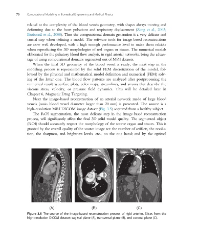
Next the image-based reconstruction of an arterial network made of large blood
vessels (main blood vessel diameter larger than 20 mm) is presented. The source is a
high-resolution MRI DICOM image dataset (Fig. 3.5) acquired from a healthy subject.
The ROI segmentation, the most delicate step in the image-based reconstruction
process, will significantly affect the final 3D solid model quality. The segmented object
(ROI) should accurately respect the morphology of the source organ and tissues. This is
granted by the overall quality of the source image set: the number of artifacts, the resolu-
tion, the sharpness, and brightness levels, etc., on the one hand; and by the optimal
(A) (B) (C)
Figure 3.5 The source of the image-based reconstruction process of rigid arteries. Slices from the
high-resolution DICOM dataset: sagittal plane (A), transversal plane (B), and coronal plane (C).