
Page 91 - Computational Modeling in Biomedical Engineering and Medical Physics
P. 91
Computational domains 79
(A) (B)
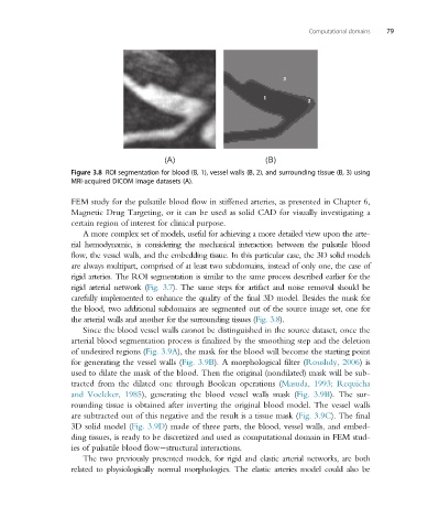
Figure 3.8 ROI segmentation for blood (B, 1), vessel walls (B, 2), and surrounding tissue (B, 3) using
MRI-acquired DICOM image datasets (A).
FEM study for the pulsatile blood flow in stiffened arteries, as presented in Chapter 6,
Magnetic Drug Targeting, or it can be used as solid CAD for visually investigating a
certain region of interest for clinical purpose.
A more complex set of models, useful for achieving a more detailed view upon the arte-
rial hemodynamic, is considering the mechanical interaction between the pulsatile blood
flow, the vessel walls, and the embedding tissue. In this particular case, the 3D solid models
are always multipart, comprised of at least two subdomains, instead of only one, the case of
rigid arteries. The ROI segmentation is similar to the same process described earlier for the
rigid arterial network (Fig. 3.7). The same steps for artifact and noise removal should be
carefully implemented to enhance the quality of the final 3D model. Besides the mask for
the blood, two additional subdomains are segmented out of the source image set, one for
the arterial walls and another for the surrounding tissues (Fig. 3.8).
Since the blood vessel walls cannot be distinguished in the source dataset, once the
arterial blood segmentation process is finalized by the smoothing step and the deletion
of undesired regions (Fig. 3.9A), the mask for the blood will become the starting point
for generating the vessel walls (Fig. 3.9B). A morphological filter (Roushdy, 2006)is
used to dilate the mask of the blood. Then the original (nondilated) mask will be sub-
tracted from the dilated one through Boolean operations (Masuda, 1993; Requicha
and Voelcker, 1985), generating the blood vessel walls mask (Fig. 3.9B). The sur-
rounding tissue is obtained after inverting the original blood model. The vessel walls
are subtracted out of this negative and the result is a tissue mask (Fig. 3.9C). The final
3D solid model (Fig. 3.9D) made of three parts, the blood, vessel walls, and embed-
ding tissues, is ready to be discretized and used as computational domain in FEM stud-
ies of pulsatile blood flow structural interactions.
The two previously presented models, for rigid and elastic arterial networks, are both
related to physiologically normal morphologies. The elastic arteries model could also be