
Page 89 - Computational Modeling in Biomedical Engineering and Medical Physics
P. 89
Computational domains 77
(A) (B) (C)
(D) (E)
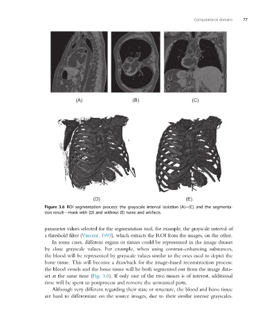
Figure 3.6 ROI segmentation process: the grayscale interval isolation (A) (C) and the segmenta-
tion result—mask with (D) and without (E) noise and artifacts.
parameter values selected for the segmentation tool, for example, the grayscale interval of
a threshold filter (Vincent, 1993), which extracts the ROI from the images, on the other.
In some cases, different organs or tissues could be represented in the image dataset
by close grayscale values. For example, when using contrast-enhancing substances,
the blood will be represented by grayscale values similar to the ones used to depict the
bone tissue. This will become a drawback for the image-based reconstruction process:
the blood vessels and the bone tissue will be both segmented out from the image data-
set at the same time (Fig. 3.6). If only one of the two tissues is of interest, additional
time will be spent to postprocess and remove the unwanted parts.
Although very different regarding their state or structure, the blood and bone tissue
are hard to differentiate on the source images, due to their similar intense grayscales.