
Page 86 - Computational Modeling in Biomedical Engineering and Medical Physics
P. 86
74 Computational Modeling in Biomedical Engineering and Medical Physics
tomography (CT) or magnetic resonance imaging (MRI) image-based reconstructed
solid models (Jan, 2006) are used as computational domains to numerically simulate
physical processes such as hemodynamic flows, with and without interaction with the
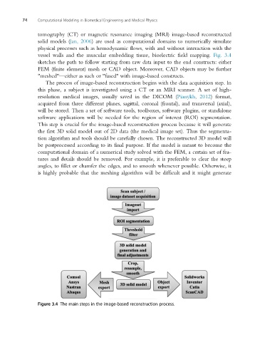
vessel walls and the muscular embedding tissue, bioelectric field mapping. Fig. 3.4
sketches the path to follow starting from raw data input to the end constructs: either
FEM (finite element) mesh or CAD object. Moreover, CAD objects may be further
"meshed"—either as such or "fused" with image-based constructs.
The process of image-based reconstruction begins with the data acquisition step. In
this phase, a subject is investigated using a CT or an MRI scanner. A set of high-
resolution medical images, usually saved in the DICOM (Pianykh, 2012) format,
acquired from three different planes, sagittal, coronal (frontal), and transversal (axial),
will be stored. Then a set of software tools, toolboxes, software plugins, or standalone
software applications will be needed for the region of interest (ROI) segmentation.
This step is crucial for the image-based reconstruction process because it will generate
the first 3D solid model out of 2D data (the medical image set). Thus the segmenta-
tion algorithm and tools should be carefully chosen. The reconstructed 3D model will
be postprocessed according to its final purpose. If the model is meant to become the
computational domain of a numerical study solved with the FEM, a certain set of fea-
tures and details should be removed. For example, it is preferable to clear the steep
angles, to fillet or chamfer the edges, and to smooth whenever possible. Otherwise, it
is highly probable that the meshing algorithm will be difficult and it might generate
Figure 3.4 The main steps in the image-based reconstruction process.