
Page 208 - Computational Retinal Image Analysis
P. 208
204 CHAPTER 11 Structure-preserving guided retinal image filtering
processing algorithms, etc. One factor that has been overlooked by most researchers
is the presence of disease. Our experience shows that some diseases might affect the
imaging of the retina. In fundus imaging, the illumination light passes through the
lens of the human eye before reaching the retina, where it is reflected back to the
camera to form the image. However, the human lens is not a perfect optical system
and it often attenuates the light passing along the path. The attenuation can be serious
when the lens is affected by diseases such as cataracts. Cataract leads to the clouding
of the lens, which implies attenuation and scattering of the light travelling through it.
This is similar to the case of a cloudy camera lens reducing the quality of a picture.
We refer to this as clouding and to the processing to remove the effect as declouding.
Studies show that cataract accounts for 33% of blindness worldwide [47] and its
global prevalence in adults over 50 years of age was about 47.8% in 2002 [48]. The
high prevalence of the disease makes it an important factor that cannot be neglected.
The retinal images are often degraded at different levels of severity, depending on
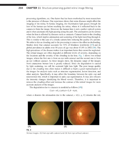
the locations and the severity of the clouding in the lens. Fig. 3 shows two retinal
images, where the first one is from an eye with cataract and the second one is from
an eye without cataract. As these images show, the dynamic range of the images
from cataractous human lens is greatly reduced. Since the degradation is caused
by light scattering, we call the scattered light lens light. The poor image quality
due to the clouding lens often makes it difficult to learn a good representation of
the images for analysis tasks such as structure segmentation, lesion detection, and
other analysis. Specifically, it may affect the boundary between the optic cup and
neuroretinal rim, which is important in optic cup segmentation. It may also obscure
the intensity changes identifying the blood vessels. Therefore, it is important to
remove the clouding effect and increase the contrast of the retinal images for more
accurate analysis of the images.
The degradation due to cataracts is modeled as follows [49]:
I p() = α L rp tp() + L ( − t p)), (1)
()
1
(
c c
c
c
where α denotes the attenuation due to the cataract; c ∈{r, g, b} denotes the red,
(A) (B)
FIG. 3
Retinal images: (A) from eye with cataract and (B) from eye without cataract.