
Page 207 - Computational Retinal Image Analysis
P. 207
1 Introduction 203
Inverse Polar Transformation Output Up-Sample Down-Sample MaxPooling DeConv <2x2, 2> Conv <1x1, 1>, with Sigmoid Conv <3x3, 1>, with ReLU Copy and Merge Multi-label Loss
+ 400 x 400 Side-Output + Multi-label
Multi-label Map L (1) s L (2) s L (3) s L (4) s
2 2 2 2
32 400 x 400 400 x 400 400 x 400
32
64
64 400 x 400 64
Segmentation 128 128
M-Net 200 x 200 128
256 256
256 512
100 x 100
Transformation 50 x 50 512 U-Shape Convolutional Network
Polar 512
256
256 25 x 25
Optic Disc Detection 128 256 384
128
64 192
64
96
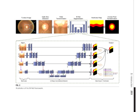
32 32 Illustration of the M-Net framework.
Fundus Image Input 3 64 3 128 3 256 Multi-scale
3
FIG. 2
50 x 50
100 x 100
400 x 400
200 x 200