
Page 214 - Computational Retinal Image Analysis
P. 214
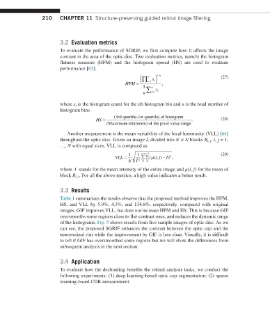
210    CHAPTER 11  Structure-preserving guided retinal image filtering


                         3.2  Evaluation metrics
                         To evaluate the performance of SGRIF, we first compute how it affects the image
                         contrast in the area of the optic disc. Two evaluation metrics, namely the histogram
                         flatness measure (HFM) and the histogram spread (HS) are used to evaluate
                         performance [63]:

                                                            i (∏ n  x i) 1/ n            (27)
                                                   HFM =    =1    ,
                                                          1 ∑ n  x
                                                          n  i=1  i

                         where x i  is the histogram count for the ith histogram bin and n is the total number of
                         histogram bins.
                                                                )
                                               (3rd quartile-1st quartile of histogram
                                       HS =                                   .          (28)
                                            (Maximum-minimum)) of the pixel value range
                            Another measurement is the mean variability of the local luminosity (VLL) [64]
                         throughout the optic disc. Given an image I, divided into N × N blocks B i, j , i, j = 1,
                         …, N with equal sizes. VLL is computed as
                                                    1   1  i=1  j=1  −                   (29)
                                                                     2
                                                             µ
                                               VLL =     ∑ ∑(( ij − ),
                                                                ,)
                                                                   I
                                                    N  I  2  N N
                               −
                         where  I  stands for the mean intensity of the entire image and μ(i, j) for the mean of
                         block B i, j . For all the above metrics, a high value indicates a better result.
                         3.3  Results
                         Table 1 summarizes the results observe that the proposed method improves the HFM,
                         HS,  and VLL  by  5.9%,  4.3%,  and  134.8%,  respectively,  compared  with  original
                         images. GIF improves VLL, but does not increase HFM and HS. This is because GIF
                         oversmooths some regions close to flat-contrast ones, and reduces the dynamic range
                         of the histograms. Fig. 5 shows results from five sample images of optic disc. As we
                         can see, the proposed SGRIF enhances the contrast between the optic cup and the
                         neuroretinal rim while the improvement by GIF is less clear. Visually, it is difficult
                         to tell if GIF has oversmoothed some regions but we will show the differences from
                         subsequent analysis in the next section.

                         3.4  Application
                         To evaluate how the declouding benefits the retinal analysis tasks, we conduct the
                         following experiments: (1) deep learning-based optic cup segmentation; (2) sparse
                         learning-based CDR measurement.