
Page 230 - Computational Retinal Image Analysis
P. 230
3 Type of lesions/clinical features 227
FIG. 4
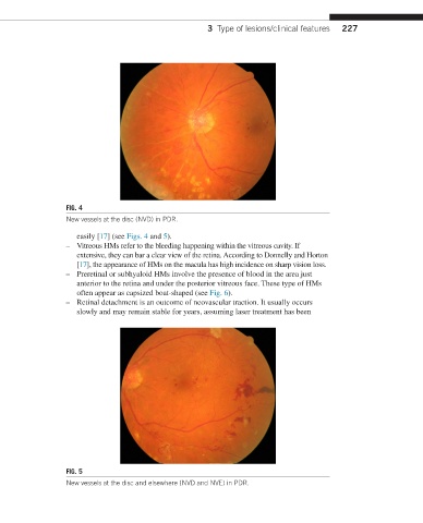
New vessels at the disc (NVD) in PDR.
easily [17] (see Figs. 4 and 5).
– Vitreous HMs refer to the bleeding happening within the vitreous cavity. If
extensive, they can bar a clear view of the retina. According to Donnelly and Horton
[17], the appearance of HMs on the macula has high incidence on sharp vision loss.
– Preretinal or subhyaloid HMs involve the presence of blood in the area just
anterior to the retina and under the posterior vitreous face. These type of HMs
often appear as capsized boat-shaped (see Fig. 6).
– Retinal detachment is an outcome of neovascular traction. It usually occurs
slowly and may remain stable for years, assuming laser treatment has been
FIG. 5
New vessels at the disc and elsewhere (NVD and NVE) in PDR.