
Page 231 - Computational Retinal Image Analysis
P. 231
228 CHAPTER 12 Diabetic retinopathy and maculopathy lesions
FIG. 6
Subhyaloid HM secondary to PDR.
applied to control the neovascular process [17].
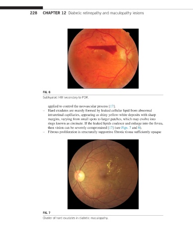
– Hard exudates are mainly formed by leaked cellular lipid from abnormal
intraretinal capillaries, appearing as shiny yellow-white deposits with sharp
margins, varying from small spots to larger patches, which may evolve into
rings known as circinate. If the leaked lipids coalesce and enlarge into the fovea,
then vision can be severely compromised [17] (see Figs. 7 and 8).
– Fibrous proliferation is structurally supportive fibrotic tissue sufficiently opaque
FIG. 7
Cluster of hard exudates in diabetic maculopathy.