
Page 236 - Computational Retinal Image Analysis
P. 236
4 Lesion detection and segmentation 233
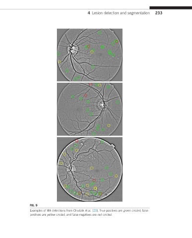
FIG. 9
Examples of MA detections from Chudzik et al. [20]. True positives are green circled, false
positives are yellow circled, and false negatives are red circled.