
Page 232 - Computational Retinal Image Analysis
P. 232
4 Lesion detection and segmentation 229
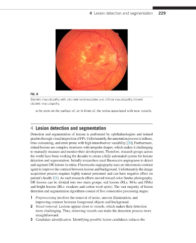
FIG. 8
Diabetic maculopathy with circinate hard exudates and diffuse maculopathy (mixed
diabetic maculopathy.
to be seen on the surface of, or in front of, the retina associated with new vessels.
4 Lesion detection and segmentation
Detection and segmentation of lesions is performed by ophthalmologists and trained
graders through visual inspection of FPs. Unfortunately, the annotation process is tedious,
time-consuming, and error-prone with high interobserver variability [20]. Furthermore,
retinal lesions are complex structures with irregular shapes, which makes it challenging
to manually measure and monitor their development. Therefore, research groups across
the world have been working for decades to create a fully automated system for lesions
detection and segmentation. Initially researchers used fluorescein angiograms to detect
and segment DR lesions in retina. Fluorescein angiography uses an intravenous contrast
agent to improve the contrast between lesions and background. Unfortunately the image
acquisition process requires highly trained personnel and can have negative effect on
patient’s health [21]. As such research efforts moved toward color fundus photography.
DR lesions can be divided into two main groups: red lesions (RLs: MAs and HMs)
and bright lesions (BLs: exudates and cotton wool spots). The vast majority of lesion
detection and segmentation algorithms consist of five consecutive processing stages:
1 Preprocessing involves the removal of noise, uneven illumination, and
improving contrast between foreground objects and background.
2 Vessel removal. Lesions appear close to vessels, which makes their detection
more challenging. Thus, removing vessels can make the detection process more
straightforward.
3 Candidate identification. Identifying possible lesion candidates reduces the