
Page 246 - Academic Press Encyclopedia of Physical Science and Technology 3rd Analytical Chemistry
P. 246
P1: GRB/GWT P2: GPJ/GAX QC: GAE/FYD Final Pages
Encyclopedia of Physical Science and Technology EN008M-395 June 29, 2001 15:52
Magnetic Resonance in Medicine 971
In MR spectroscopy, the molecules being studied are
present in a concentration very low compared to that of
water. As a consequence, spectroscopy signals tend to
be very weak. This places an even stronger premium on
field strength than does imaging. It is widely accepted that
spectroscopic studies of patients are not warranted in field
strengths less than about 1.5 T.
B. Magnets
The magnet is probably the most significant portion of a
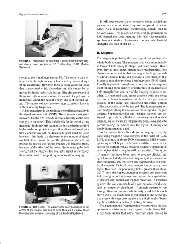
FIGURE 4 Preparation for scanning. The superconducting mag-
whole-body scanner. The magnets used vary substantially
net shown here operates at 1.5 T. (Courtesy of GE Medical
in terms of field strength, shape, and basic design. How-
Systems.)
ever, they all must meet certain basic requirements. One
obvious requirement is that the magnet be large enough
2
strength, the signal increases as B . The noise in the sys- to admit a human body and produce a field strength that
0
tem can be brought to a very low level by proper design is intense enough to produce a strong proton NMR signal.
of the electronics. However, there is some electrical noise Equally important, though not so obvios, is the require-
that is generated within the patient and that cannot be re- ment for high homogeneity, or uniformity of the magnetic
moved by improved circuit design. The ultimate source of field strength from one part of the imaging volume to an-
this noise is the random motion of ions and charged macro- other. It is essential that, unless gradient coils are being
molecules within the patient’s body and is of thermal ori- used to deliberately modulate it, the Larmor precession
gin. The noise voltage increases approximately linearly proceed at the same rate throughout the entire portion
with increasing frequency. of the patient that is to be imaged. The homogeneity re-
A key parameter in determining overall image quality is quirment puts strong limitations on the acceptable magnet
the signal-to-noise ratio (SNR). The arguments just given designs. Superconducting and resistive systems are de-
indicate that the SNR should increase linearly as the field signed to provide a cylindrical symmetry. A cylindrical
strength is increased. This is the basis for the use of strong opening, called the room temperature bore, is available to
magnetic fields in NMR scanners. If it is desired to achieve permit placing the patient into the region of strong and
high-resolution proton images, thin slices and small pic- highly homogeneous field.
ture elements (as will be discussed later) must be used; At the present time clinical proton imaging is usually
however, this leads to a decrease in the amount of signal done using magnetic field strengths in the order of 0.2 to
available to determine the pixel brightness numbers. If this 1.5 T, although, in about 2000, commercial MRI systems
process is pushed too far, the images will become grainy operating at3Tbegan to become available. Later, in the
because of the effects of the noise. By increasing the field section on current trends, research scanners operating at
strength of the magnet, the available signal is increased; even higher field strengths will be described. The types
this can be used to support higher resolution imaging. of magnet that have been used to produce clinical im-
ages have included permanent magnet systems, iron-core
electromagnets, and resistive and superconducting mul-
ticoil magnets. Each of these designs has certain advan-
tages. However, for producing fields greater than about
0.5 T, only the superconducting systems are practical;
field strengths in this range are beyond the capabilities
of present-day permanent magnet materials. In resistive
systems the coils are made of a conventional conductor,
such as copper or aluminum. If enough current is run
through them to produce whole-body sized fields much
above 0.2 T, so much heat is generated in the windings
that even with water cooling there is a likelihood of burn-
ing the insulation or actually melting the wire.
Thephenomenonofsuperconductivityprovidesthebest
FIGURE 5 NMR scan. The patient has been positioned in the
center of the magnet and can be seen through a window above approach to achieving strong magnetic fields. Since 1911
the operator’s console. (Courtesy of GE Medical Systems.) it has been known that some materials when cooled to