
Page 176 - Academic Press Encyclopedia of Physical Science and Technology 3rd BioChemistry
P. 176
P1: GPAFinal Pages
Encyclopedia of Physical Science and Technology EN013D-616 July 27, 2001 12:5
216 Protein Structure
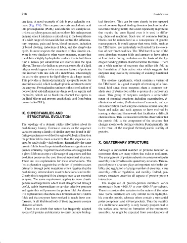
one face. A good example of this is prostaglandin syn- ical functions. This can be seen clearly in the repeated
thase (Fig. 17d). This enzyme converts arachidonic acid use of common ligand binding domains (such as the din-
into prostaglandin (PGH 2 ) and exhibits two catalytic ac- ucleotide binding motif) that occur repeatedly in proteins
tivities: a cycloxygenase and peroxidase. It is an important that require the same ligand even it is used in differ-
enzyme since it catalyzes a critical step in the biosynthesis ing chemical reactions. Such use of common building
of a wide range of eicosanoids that control the inflamma- blocks can be rationalized as a consequence of genetic
tory response, pain and fever, blood pressure, induction rearrangement. It would appear that some folds, such as
of blood clotting, induction of labor, and the sleep/wake the TIM barrel, are particularly well suited for the evolu-
cycle. In most respects the structure of this dimeric en- tion of new functionalities. The TIM barrel is one of the
zyme is very similar to other water enzymes, except that most abundant enzyme folds and appears to have arisen
it exhibits a highly hydrophobic surface that is built from at least twice during evolution on the basis of the hy-
four α-helices per subunit that are inserted into the lipid drogen bonding pattern observed within the barrel. There
bilayer. The use of α-helices to penetrate one side of a lipid are a wide number of enzymes that utilize this fold as
bilayer would appear to be a common feature of proteins the foundation of their active sites which suggests that
that interact with one side of a membrane. Interestingly enzymes may evolve by retooling of existing functional
the active site opens to the lipid bilayer via a large tunnel. folds.
This provides a thermodynamically acceptable route for The enolase superfamily, which contains a variant of
arachidonic acid, which is a hydrophobic substrate to enter the TIM barrel, is a good example of retooling of a func-
the enzyme. Prostaglandin synthase is the site of action of tional fold since these enzymes share a common cat-
nonsteroidal anti-inflammatory drugs such as aspirin and alytic step of abstraction of the α-proton of a carboxylate
ibuprofen, which act to block the channel that opens to anion. This group of enzymes catalyzes a remarkable
the lipid bilayer and prevent arachidonic acid from being range of chemical reactions including racemization, β-
converted to PGH 2 . elimination of water, β-elimination of ammonia, and cy-
cloisomerization. Each enzyme contains similar catalytic
bases and acids and each appears to have evolved by
IX. SUPERFAMILIES AND reusing a structural framework that facilitates a difficult
STRUCTURAL EVOLUTION chemical task. This is consistent with the observation that
the protein fold is the component of the structure that
The topology of a domain yields information about its changes most slowly during evolution. Fundamentally this
evolutionary history. Extensive studies on the sequence is the result of the marginal thermodynamic stablity of
variation among a family of similar enzymes found in dif- proteins.
fering organisms reveal that for a given biological function
the protein fold is more conserved than the sequence, ex-
cept for catalytically vital residues. Remarkably the same X. QUATERNARY STRUCTURE
proteinfoldisfoundinproteinsthatsharenosignificantse-
quencesimilarity.Togethertheseobservationssuggestthat Although a substantial number of proteins function as
a given fold can accept a wide range of sequences and that monomers there are many others that exist as multimers.
evolution preserves the core three-dimensional structure. The arrangement of protein subunits in a macromolecular
There are two explanations for these observations. The assembly is referred to as its quaternary structure. This as-
first explanation suggests that evolution of proteins occurs pect of protein structure plays an important role in the sta-
primarily through point mutations which requires that the bility and regulation of a large number of enzymes, virus
evolutionary intermediates must be functional and stable. assembly, cellular regulation, and motility. Indeed, qua-
Clearly this is required if the changes involve an essential ternary structure underlies all aspects of protein–protein
enzyme. The same requirement applies to more drastic interaction.
genetic rearrangements which must also proceed through The magnitude of protein–protein interfaces varies
˚ 2
˚ 2
useful, stable intermediates to survive selective pressure enormously from ∼800 A to over 4000 A per subunit.
and again this will preserve the protein fold. An alterna- There is considerable variation in the nature of the inter-
tive explanation is that there are a limited number of stable face. Some interfaces are very similar to the hydropho-
folds and that enzymes have evolved to reach these con- bic core the protein, whereas others contain a substantial
formers. In all likelihood both of these arguments contain polar component and solvent pockets. Thus the stability
elements of truth. of a multimeric assembly is only loosely proportional to
There is no doubt that nature has frequently adapted the surface area buried on formation of the multimeric
successful protein architectures to carry out new biolog- assembly. As might be expected from considerations of