
Page 151 - Environmental Nanotechnology Applications and Impacts of Nanomaterials
P. 151
Methods for Structural and Chemical Characterization of Nanomaterials 137
ratio of the ellipsoid and agglomeration stage. Even slightly agglomer-
ated nanoparticles would have displayed a strong increase of the scat-
tering intensity in the low q region. In summary, using small angle
X-ray scattering, information on the size, shape, and interaction between
nanoparticles in solution can be obtained, even at high concentration
suspensions. However, this method is not well-adapted for characteri-
zation of complex mixtures or very small nanoparticle concentration.
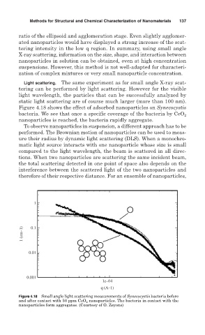
Light scattering. The same experiment as for small angle X-ray scat-
tering can be performed by light scattering. However for the visible
light wavelength, the particles that can be successfully analyzed by
static light scattering are of course much larger (more than 100 nm).
Figure 4.18 shows the effect of adsorbed nanoparticles on Synecocystis
bacteria. We see that once a specific coverage of the bacteria by CeO 2
nanoparticles is reached, the bacteria rapidly aggregate.
To observe nanoparticles in suspension, a different approach has to be
performed. The Brownian motion of nanoparticles can be used to meas-
ure their radius by dynamic light scattering (DLS). When a monochro-
matic light source interacts with one nanoparticle whose size is small
compared to the light wavelength, the beam is scattered in all direc-
tions. When two nanoparticles are scattering the same incident beam,
the total scattering detected in one point of space also depends on the
interference between the scattered light of the two nanoparticles and
therefore of their respective distance. For an ensemble of nanoparticles,
1
1 (cm–1) 0.1
0.01
0.001
1e–04
q (A–1)
Figure 4.18 Small angle light scattering measurements of Synecocystis bacteria before
and after contact with 50 ppm CeO 2 nanoparticles. The bacteria in contact with the
nanoparticles form aggregates. (Courtesy of O. Zeyons)