Page 239 - Essentials of physical chemistry
P. 239
Basic Spectroscopy                                                          201

            windings in a transformer to create a higher voltage because you need alternating current to increase
            voltage with a transformer. We also show the use of a simple diode to allow a half-wave rectification
            of the high voltage alternating current to create a pulsed direct current beam. That is only for an
            educational schematic, in an actual device there would be full-wave rectification with capacitors
            in parallel to damp ripples in the direct current to a high degree so as to allow a continuous electron
            beam to approach the optical properties of a microscope using visible light. The idea of an electron
            microscope is to use a beam of electrons in place of a light beam. Thus, an electron microscope uses
            very sophisticated electrostatic lenses to simulate optical lenses so the diagram is very crude but
            emphasizes the Auger process and the dispersal of K a or L a x-rays into a range of characteristic
            bands for the various elements.
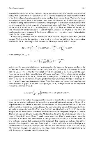
              Let us develop a formula from the Bohr model, which shows the way to calculate the K a for each
            element. We know the K a transition is from n ¼ 2to n ¼ 1, so we will have the same quantum
            numbers for the K a wavelength of all elements (in this simple model), only Z changes.


                                    1   1               12398    2  3
                                2
                         DE ¼ (Z )         (13:6057 eV) ¼    ¼ Z     (13:6057)
                                   1 2  2 2              l K a     4
                               as
            so we rearrange for l K a

                                             12, 398   4     1214:981
                                  l K a  (A ˚ ) ¼          ¼
                                            13:6057   3Z 2      Z  2
            and we see the wavelength is inversely proportional to the square of the atomic number of the
            element. Thus, if we want to calculate the wavelength of the K a wavelength for cadmium we would
            find for Cd, Z ¼ 48, so that the wavelength would be estimated as 0.527335 Å for the x-ray.
            However, we saw the Bohr model led to a 6.2% error for Cu and Cd has a larger atomic number.
            The experimental value for the K a fluorescence wavelength of Cd is 0.5357 Å with only a 1.6%
            error, so we see our simple Bohr model is good for the heavier elements. In order to eliminate this
            error it would be necessary to run experimental Auger fluorescence on known samples and create a
            correction table or graph. If we want to compute just the K a voltage we can use the expression
            reduced to a constant times Z 2


                                      1   1                 2
                                   2
                            DE ¼ (Z )        (13:6057 eV) ¼ (Z )(10:204275 eV):
                                      1 2  2 2
            In the opinion of this author, it is important for students to learn the basic principles of instrumen-
            tation but we need an application for motivation so an actual spectrum is shown in Figure 9.9 as
            output obtained for a sample of lead shot. It is evident that the lead is in abundance but is not pure
            and several other elements are present. Perhaps the most interesting feature of the scan is that there
            are really two peaks for lead, M a (2.343 keV, 5.076 Å) and M b (2.442 keV, 5.299 Å) that are the
            energies emitted when the first and second electrons fall down into the partially empty n ¼ 3 orbital.
            The less intense peak at higher energy is the M b transition. Note that the entire spectrum falls with a
            range of (0–10 keV), so the K and L transitions are not reported but M transitions are within the
            parameters of the instrument. The detection of small peaks for other elements is treated with the aid
            of a software program that accompanies the instrument. For forensic applications, the relative
            amounts of the impurities could vary from sample to sample and might be matched uniquely to
            some unfired shot in a box of cartridges=shells obtained from a suspect in a crime. Thus, this sample
            XRF spectrum illustrates M a,b transitions for a heavy metal of interest to forensic problems along
            with illustration of elemental impurities in the lead.

