
Page 249 - Fiber Fracture
P. 249
STRENGTH AND FRACTURE OF METALLIC FILAMENTS 233
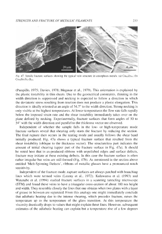
Fig. 47. Tensile fracture surfaces showing the typical vein structure in amorphous metals: (a) Cu.joZr.jn; (b)
Co7nFesSi isB in.
(Pampillo, 1975; Davies, 1978; Megusar et al., 1979). This orientation is explained by
the plastic instability in thin sheets. Due to the geometrical constraints, thinning in the
width direction is suppressed and necking is expected to follow a direction in which
the deviatoric stress resulting from traction does not produce a plastic elongation. This
direction is ideally oriented at an angle of 54.7" to the width direction. Strong necking is
only visible at the highest temperatures. At lower temperatures the flow rate falls rapidly
below the imposed strain rate and the shear instability immediately takes over on the
plane defined by necking. Experimentally, fracture surfaces that form angles of 50 to
54" with the width direction and parallel to the thickness vector are observed.
Independent of whether the sample fails in the low- or high-temperature mode
fracture surfaces reveal that shearing only starts the fracture by reducing the section.
The final rupture then occurs in the tearing mode and usually follows the shear band
initially produced. Fig. 47a shows a typical fracture surface that resulted from the
shear instability (oblique to the thickness vector). The structureless part indicates the
amount of initial shearing (upper part of the fracture surface in Fig. 47a). It should
be noted here that in as-produced ribbons with unpolished edges and surface defects,
fracture may initiate at these existing defects. In this case the fracture surface is often
rather irregular but veins are still formed (Fig. 47b). As mentioned in the section above
entitled 'Melt-Spinning Defects', ribbons of metallic glasses have a pronounced notch
sensitivity.
Independent of the fracture mode, rupture surfaces are always patched with branching
lines which were termed veins (Leamy et al., 1972). Kulawansa et al. (1993) and
Watanabe et al. (1994) studied fracture surfaces in a scanning tunneling microscope
(STM) and found these veins to have a triangular cross-section of about 100 nm height
and width. They resemble closely the lines that one obtains when two plates with a layer
of grease in between are separated. From this analogy one might immediately conclude
that adiabatic heating due to the intense shearing, which precedes fracture, raises the
temperature up to the temperature of the glass transition. At this temperature the
viscosity drastically drops to values that might explain these lines. However, subsequent
estimates of the adiabatic heating can explain but a temperature rise of a few degrees