
Page 203 - Fundamentals of Light Microscopy and Electronic Imaging
P. 203
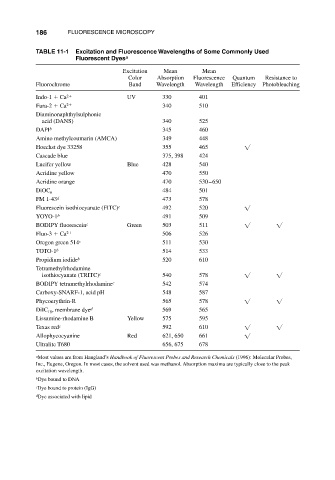
186      FLUORESCENCE MICROSCOPY

                       TABLE 11-1  Excitation and Fluorescence Wavelengths of Some Commonly Used
                                   Fluorescent Dyes a

                                                 Excitation  Mean        Mean
                                                  Color    Absorption  Fluorescence  Quantum  Resistance to
                       Fluorochrome                Band    Wavelength  Wavelength  Efficiency  Photobleaching
                       Indo-1   Ca 2              UV         330        401
                       Fura-2   Ca 2                         340        510
                       Diaminonaphthylsulphonic
                        acid (DANS)                          340        525
                       DAPI b                                345        460
                       Amino methylcoumarin (AMCA)           349        448
                       Hoechst dye 33258                     355        465
                       Cascade blue                          375, 398   424
                       Lucifer yellow             Blue       428        540
                       Acridine yellow                       470        550
                       Acridine orange                       470        530–650
                       DiOC                                  484        501
                           6
                       FM 1-43 d                             473        578
                       Fluorescein isothiocyanate (FITC) c   492        520
                       YOYO-1 b                              491        509
                       BODIPY fluorescein c       Green      503        511
                       Fluo-3   Ca 2                         506        526
                       Oregon green 514 c                    511        530
                       TOTO-1 b                              514        533
                       Propidium iodide b                    520        610
                       Tetramethylrhodamine
                        isothiocyanate (TRITC) c             540        578
                       BODIPY tetramethylrhodamine c         542        574
                       Carboxy-SNARF-1, acid pH              548        587
                       Phycoerythrin-R                       565        578
                       DiIC , membrane dye d                 569        565
                          18
                       Lissamine-rhodamine B      Yellow     575        595
                       Texas red c                           592        610
                       Allophycocyanine           Red        621, 650   661
                       Ultralite T680                        656, 675   678
                       a Most values are from Haugland’s Handbook of Fluorescent Probes and Research Chemicals (1996): Molecular Probes,
                       Inc., Eugene, Oregon. In most cases, the solvent used was methanol. Absorption maxima are typically close to the peak
                       excitation wavelength.
                       b Dye bound to DNA
                       c Dye bound to protein (IgG)
                       d Dye associated with lipid