
Page 206 - Fundamentals of Light Microscopy and Electronic Imaging
P. 206
ARRANGEMENT OF FILTERS AND THE EPI-ILLUMINATOR 189
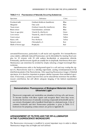
TABLE 11-2 Fluorescence of Naturally Occurring Substances
Specimen Substance Color
Powdered milk Oxidized riboflavin (lumiflavin) Blue
Margarine Fatty acids Blue
Yeast extract Oxidized vitamin B (lumiflavin) Blue
2
Brain extract Catecholamines, serotonin Blue
Yeast on agar plate Vitamin B (riboflavin) Green
2
Liver extract Vitamin B , other B vitamins Yellow
2
Carrot extract -carotene Yellow
Butter, milk Free riboflavin Yellow
Spinach extract Chlorophyll a, b Red
Shells of brown eggs Porphyrins Red
unwanted fluorescence, particularly in cell nuclei and organelles. For immunofluores-
cence studies, aldehyde-induced fluorescence can be diminished by treating fixed sam-
ples for 10 minutes with 20 mM sodium borohydride or ammonium chloride.
Fortunately, autofluorescent signals are usually low in amplitude. Interference from auto-
fluorescence can sometimes be avoided by simply selecting a longer-wavelength fluo-
rochrome.
Autofluorescence adds to the background signal in a cell and may overlap the sig-
nal of a fluorochrome used in a labeling experiment, causing misinterpretation of the
distribution pattern of the fluorochrome. After acquiring fluorescence images of labeled
specimens, it is therefore important to prepare similar exposures from unlabeled speci-
mens. If necessary, a camera exposure time can be selected that minimizes the autofluo-
rescent contribution, but still allows adequate imaging of the labeled experimental
material.
Demonstration: Fluorescence of Biological Materials Under
Ultraviolet Light
Fluorescent compounds and metabolites are abundant in living cells and tissues.
To become familiar with these signals and recognize them when they occur,
examine the fluorescence of naturally occurring compounds in foodstuffs and tis-
sue extracts illuminated with a handheld black light in a darkened room. A list of
common foodstuffs and their fluorescence properties is given in Table 11-2.
Instructions for preparing certain extracts are given in Appendix II.
ARRANGEMENT OF FILTERS AND THE EPI-ILLUMINATOR
IN THE FLUORESCENCE MICROSCOPE
The fluorescence microscope is modified in several important ways in order to obtain
fluorescence images that are bright and well defined: