
Page 207 - Fundamentals of Light Microscopy and Electronic Imaging
P. 207
190 FLUORESCENCE MICROSCOPY
• A bright light source such as a mercury or xenon arc lamp is required because only
a narrow band of wavelengths, and consequently a small portion of the illuminator
output, is used to excite fluorochromes in the specimen.
• For efficient high-contrast imaging, both the illuminator and objective lens are
positioned on the same side of the specimen. In this arrangement, the lamp and light
delivery assembly are called an epi-illuminator, and the objective lens functions
both as the condenser, delivering excitatory light to the specimen, and as the objec-
tive lens, collecting fluorescent light and forming an image of the fluorescent object
in the image plane.
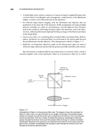
• Fluorescence filter sets containing three essential filters (excitation filter, dichroic
mirror, and barrier [or emission] filter) are positioned in the optical path between
the epi-illuminator and the objective. This arrangement is shown in Figure 11-7.
• High-NA, oil immersion objectives made of low-fluorescence glass are used to
maximize light collection and provide the greatest possible resolution and contrast.
Epi-illumination is made possible by the employment of a dichroic mirror, which is
mounted together with exciter and barrier filters as a fluorescence filter set in a filter
Barrier or
emission filter
Exciter
filter Dichroic mirror
Light
source Filter cube
Objective
Object
Figure 11-7
Arrangement of filters in a fluorescence filter cube. The diagram shows the orientation of
filters in a filter cube in an epi-illuminator for an upright microscope. The excitation beam
(dotted line) passes through the exciter and is reflected by the dichroic mirror and directed
toward the specimen. The return beam of emitted fluorescence wavelengths (solid line)
passes through the dichroic mirror and the emission filter to the eye or camera. Excitation
wavelengths reflected at the specimen are reflected by the dichroic mirror back toward the
light source. Excitation wavelengths that manage to pass through the dichroic mirror are
blocked by the barrier (emission) filter.