
Page 49 - Handbook of Properties of Textile and Technical Fibres
P. 49
30 Handbook of Properties of Textile and Technical Fibres
result in the creation of volatile species that can produce surface craters and roughness.
For fibers that are poor thermal conductors, the differential thermal expansion between
the polymer and the metallic coating can create surface cracks and blistering.
Recent progress in SEM techniques allows reduced electron impact energies with
low vacuum modes to be used. Nowadays, most tabletop SEMs provide very sensitive
low-vacuum electron detectors able to reveal the smallest details of the fiber surface.
For the most delicate samples, the use of air or atmospheric scanning electron micro-
scope have also been proposed (Suga et al., 2014). More recently, Solomonov et al.
(2014) have, for example, used an AirSEMTM equipment to image collagen fibrils
with very simplified sample preparation protocols.
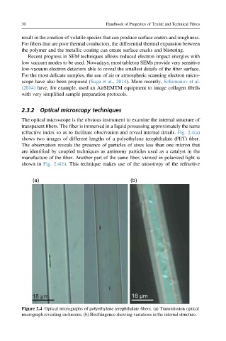
2.3.2 Optical microscopy techniques
The optical microscope is the obvious instrument to examine the internal structure of
transparent fibers. The fiber is immersed in a liquid possessing approximately the same
refractive index so as to facilitate observation and reveal internal details. Fig. 2.4(a)
shows two images of different lengths of a polyethylene terephthalate (PET) fiber.
The observation reveals the presence of particles of sizes less than one micron that
are identified by coupled techniques as antimony particles used as a catalyst in the
manufacture of the fiber. Another part of the same fiber, viewed in polarized light is
shown in Fig. 2.4(b). This technique makes use of the anisotropy of the refractive
(a) (b)
Figure 2.4 Optical micrographs of polyethylene terephthalate fibers. (a) Transmission optical
micrograph revealing inclusions. (b) Birefringence showing variations in the internal structure.