
Page 50 - Handbook of Properties of Textile and Technical Fibres
P. 50
Testing and characterization of fibers 31
index of the fiber so that the effect of birefringence occurs and can be used to reveal
variations in the degree of orientation of the fiber internal structure and may demon-
strate the existence of local structural variations such as in skin/core properties.
More quantitative insights on the degree of orientation of the fiber can be obtained
by measuring optical birefringence using a Berek compensator and can be related to
other coupled methods such as X-ray diffraction and infrared dichroism (Stein and
Norris, 1956).
Microtomy is a technique developed first for histology, the study of biological ma-
terials. By the use of a fine knife or blade the material is cut into thin slices with thick-
nesses less than 5 mm, and an ultramicrotome is used to obtain thickness of around 1
micron for optical microscopy and down to 50 nm for transmission electron micro-
scopy. For the examination of fibers the specimens are usually embedded in a resin,
which is then presented to the glass or diamond knife and successive slices cut. The
knife advances at a controlled rate with respect to the specimen so that successive sec-
tions of the fiber are cut. These sections fall into water from which they are recovered
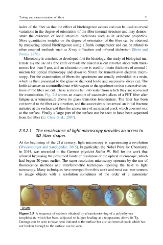
for examination. Fig. 2.5 shows an example of successive slices of a PET fiber after
fatigue at a temperature above its glass transition temperature. The fiber has been
cut normal to the fiber axis direction, and the successive slices reveal an initial fracture
initiated at the surface and then the appearance of an internal crack which does not exit
at the surface. Finally a large part of the surface can be seen to have been separated
from the fiber (Le Clerc et al., 2007).
2.3.2.1 The renaissance of light microscopy provides an access to
3D fiber shapes
At the beginning of the 21st century, light microscopy is experiencing a revolution
(Weisenburger and Sandoghdar, 2015). In particular, the Nobel Prize for Chemistry,
in 2014, was rewarded to the German physicist Stefan W. Hell for the work that
allowed bypassing the presumed limits of resolution of the optical microscope, which
had begun 20 years earlier. The super-resolution microscopy operates by the use of
fluorescence methods and interferometric techniques opening the field to light
nanoscopy. Many techniques have emerged from this work and most use laser sources
to image objects with a resolution sometimes of the order of a nanometer
10 μm
Figure 2.5 A sequence of sections obtained by ultramicrotoming of a polyethylene
terephthalate which has been subjected to fatigue loading at a temperature above its Tg.
Damage can be seen to have been initiated at the surface but also an internal crack which has
not broken through to the surface can be seen.