
Page 277 - Microtectonics
P. 277
270 10 · Special Techniques
Fig. 10.5. Polished sample of ultramylonite, etched with HF vapour. Secondary electron mode. Gold cover. Fine detail is visible through
the presence of etch pits and different grey-tones for the three minerals present. Quartz has not been etched much and forms the matrix
(light grey). Feldspar and biotite have been etched. Feldspar is dark grey and forms the central porphyroclast. The etched medium grey
elongate grains with cleavage are biotite. Biotite seems to replace feldspar at the left-hand side of the porphyroclast. St. Barthélemy
Massif, Pyrenees, France. Width of view 40 µm
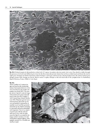
Fig. 10.6.
Atomic number or Z contrast im-
ages are based on the relationship
between backscattered electron emis-
sion characteristics and sample com-
position (e.g. Lloyd 1987). The exam-
ple shows a typical Z contrast image
obtained from low-grade metasedi-
ments of the Clew Bay Complex, S.
Achill Island, Mayo, Ireland (Yardley
et al. 1996). Polyphase garnets have a
detrital pyrope-almandine (py-al)
core surrounded by spessartine-al-
mandine (sp-al) and then grossular-
almandine (gr-al), which is unmixing
to a grossular-rich and an almandine-
rich component. They sit in a matrix
of quartz (qtz), chlorite (chl) and
white mica (mus), with the latter
zoned for Mg+Fe as revealed by the
image contrast variations (Fe-rich
compositions appear brighter). (Pho-
tograph courtesy Eric Condliffe and
Geoffrey Lloyd)