
Page 278 - Microtectonics
P. 278
10.2 · Techniques to Study Deformation Fabrics 271
which scatter at a small angle from lattice planes in a in this way are known as electron channelling (EC)
crystal. Such a diffraction pattern carries all informa- patterns and have a spatial resolution of >1–10 µm
tion necessary to determine the exact orientation of a but an angular resolution of <1°. The angular spread
crystal with an error of less than 1°, provided the type of EC patterns is typically <20°, which makes identi-
and composition of the mineral is known (Krieger- fication relatively difficult (Fig. 10.8a).
Lassen 1996; Prior 1999). In order to find the crystal 3b. with the sample in the position for FSE-OC (i.e.
orientation from the pattern, the lines in the pattern highly inclined), diffraction patterns can be ob-
must be indexed, i.e. the corresponding lattice planes tained simply by focusing the incident electron
should be identified. There are two different methods beam on a fixed position on the sample. The dif-
to obtain electron diffraction patterns: fraction patterns produced in this way are known
3a. with the sample in the position for EC-OC (i.e. hori- as electron backscattered diffraction (EBSD) pat-
zontal), diffraction patterns can be obtained if the terns and have a spatial resolution of 0.1–1.5 µm
incident electron beam is focused on a fixed posi- with an angular resolution of ~1°. The angular
tion on the sample and rocked back- and forward spread of EBSD patterns is typically >50°, which
(Figs. 10.3, 10.8a). The diffraction patterns produced makes identification relatively easy (Fig. 10.8b).
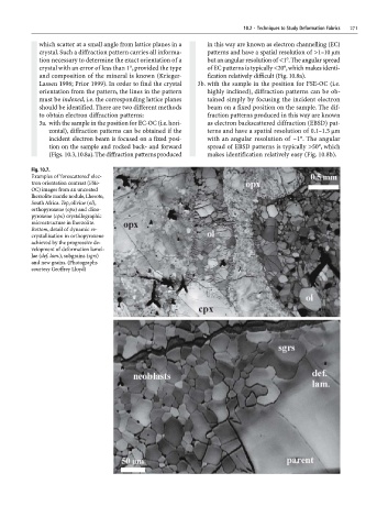
Fig. 10.7.
Examples of ‘forescattered’ elec-
tron orientation contrast (FSE-
OC) images from an uncoated
lherzolite mantle nodule, Lhesoto,
South Africa. Top, olivine (ol),
orthopyroxene (opx) and clino-
pyroxene (cpx) crystallographic
microstructure in lherzolite.
Bottom, detail of dynamic re-
crystallisation in orthopyroxene
achieved by the progressive de-
velopment of deformation lamel-
lae (def. lam.), subgrains (sgrs)
and new grains. (Photographs
courtesy Geoffrey Lloyd)