
Page 132 - Book Hosokawa Nanoparticle Technology Handbook
P. 132
FUNDAMENTALS CH. 2 STRUCTURAL CONTROL OF NANOPARTICLES
2.6.3 Design of nanoparticle surface and application 15 Dox. sol.
for DDS
Non-coated
Surface properties of fine particles affect its stability HPMC-R65
and pharmaceutical functions. When relatively soft PVA-R
particle like a liposome is mixed with water-soluble 10 * ** * **
polymer solution having a hydrophobic moiety just *
like cholesterol, its surface can be modified with the Conc. of Dox. (µg/g tumor)
polymer by penetrating the hydrophobic part of poly-
mer molecule into the lipid double layer of the lipo- 5
some (anchoring) [11]. Anchoring method is available
only for layers with high-fluid particle surface like
phospholipid layer.
For designing the novel dosage form, surface modi-
fication of liposomes was investigated in the author’s 0
laboratory. Polyvinylalcohol (PVA), which is popular 1 8 24 48
as a protective colloid, was chosen as the coating poly- Time (hr)
mer; long alkyl chain was chemically bonded at the
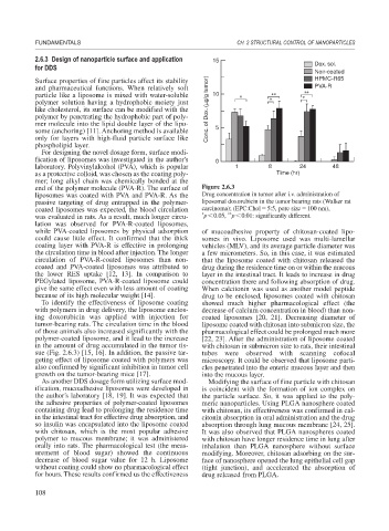
end of the polymer molecule (PVA-R). The surface of Figure 2.6.3
liposomes was coated with PVA and PVA-R. As the Drug concentration in tumor after i.v. administration of
passive targeting of drug entrapped in the polymer- liposomal doxorubicin in the tumor bearing rats (Walker rat
coated liposomes was expected, the blood circulation carcinoma); (EPC:Chol 5:5, pore size 100 nm),
**
was evaluated in rats. As a result, much longer circu- * p 0.05, p 0.01: significantly different.
lation was observed for PVA-R-coated liposomes,
while PVA-coated liposomes by physical adsorption of mucoadhesive property of chitosan-coated lipo-
could cause little effect. It confirmed that the thick somes in vivo. Liposome used was multi-lamellar
coating layer with PVA-R is effective in prolonging vehicles (MLV), and its average particle diameter was
the circulation time in blood after injection. The longer a few micrometers. So, in this case, it was estimated
circulation of PVA-R-coated liposomes than non- that the liposome coated with chitosan released the
coated and PVA-coated liposomes was attributed to drug during the residence time on or within the mucous
the lower RES uptake [12, 13]. In comparison to layer in the intestinal tract. It leads to increase in drug
PEGylated liposome, PVA-R-coated liposome could concentration there and following absorption of drug.
give the same effect even with less amount of coating When calcitonin was used as another model peptide
because of its high molecular weight [14]. drug to be enclosed, liposomes coated with chitosan
To identify the effectiveness of liposome coating showed much higher pharmacological effect (the
with polymers in drug delivery, the liposome enclos- decrease of calcium concentration in blood) than non-
ing doxorubicin was applied with injection for coated liposomes [20, 21]. Decreasing diameter of
tumor-bearing rats. The circulation time in the blood liposome coated with chitosan into submicron size, the
of those animals also increased significantly with the pharmacological effect could be prolonged much more
polymer-coated liposome, and it lead to the increase [22, 23]. After the administration of liposome coated
in the amount of drug accumulated in the tumor tis- with chitosan in submicron size to rats, their intestinal
sue (Fig. 2.6.3) [15, 16]. In addition, the passive tar- tubes were observed with scanning cofocal
geting effect of liposome coated with polymers was microscopy. It could be observed that liposome parti-
also confirmed by significant inhibition in tumor cell cles penetrated into the enteric mucous layer and then
growth on the tumor-bearing mice [17]. into the mucous layer.
As another DDS dosage form utilizing surface mod- Modifying the surface of fine particle with chitosan
ification, mucoadhesive liposomes were developed in is coincident with the formation of ion complex on
the author’s laboratory [18, 19]. It was expected that the particle surface. So, it was applied to the poly-
the adhesive properties of polymer-coated liposomes meric nanoparticles. Using PLGA nanosphere coated
containing drug lead to prolonging the residence time with chitosan, its effectiveness was confirmed in cal-
in the intestinal tract for effective drug absorption, and citonin absorption in oral administration and the drug
so insulin was encapsulated into the liposome coated absorption through lung mucous membrane [24, 25].
with chitosan, which is the most popular adhesive It was also observed that PLGA nanospheres coated
polymer to mucous membrane; it was administered with chitosan have longer residence time in lung after
orally into rats. The pharmacological test (the meas- inhalation than PLGA nanosphere without surface
urement of blood sugar) showed the continuous modifying. Moreover, chitosan adsorbing on the sur-
decrease of blood sugar value for 12 h. Liposome face of nanosphere opened the lung epithelial cell gap
without coating could show no pharmacological effect (tight junction), and accelerated the absorption of
for hours. These results confirmed us the effectiveness drug released from PLGA.
108