
Page 128 - Book Hosokawa Nanoparticle Technology Handbook
P. 128
FUNDAMENTALS CH. 2 STRUCTURAL CONTROL OF NANOPARTICLES
then tetramethoxysilane is added while stirring vigor- mesoporous silica surface. Mesoporous silica modified
ously. After several hours, precipitation occurs, and by aminopropylsilane [NH (CH ) Si(OCH CH ) ]
3 3
2
2
2 3
after ageing overnight, the solution is filtered and selectively adsorbs anionic proteins, and its release can
washed. A mesoporous material is obtained by remov- also be controlled by adjusting the pH of the solution
ing the template by calcination at 550ºC [15, 16]. [22]. Mesoporous systems that actively respond to exter-
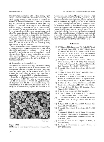
Figure 2.5.16 shows a scanning electron microscopy nal stimuli are also being formulated, making it possible
(SEM) image of mesoporous silica synthesized by to release the drug when it reaches the affected area. A
this method. The mesoporous silica shows very uni- method in which an coumarin group dimerized by irra-
form spherical morphology and monodisperse parti- diation is bonded to the pore opening has been proposed,
cles. The mean diameter of these particles is 655 nm, where light is used to control whether the pore is closed
with very little distribution in particle size (standard or open [23].Other attempts have been made to seal the
deviation: 3.7%). Moreover, with a pore diameter of pores with CdS particles, which are chemically removed
2 nm, the particles have an extremely large surface when the drug has reached the affected area [24].
2
area: approximately 1000 m /g. Mesoporous particles
from 100 nm to 3 m in size are currently being
synthesized using this method. References
In addition to the Stöber method, other techniques [1] C.T. Kresge, M.E. Leonowicz, W.J. Roth, J.C. Vartuli
for synthesizing mesoporous particles include emul- and J.S. Beck: Nature, 359, 710–712 (1992). J.S. Beck,
sion [18], and gas-phase methods [19]. Particles of J.C. Vartuli, W.J. Roth, M.E. Leonowicz, C.T. Kresge,
around 0.5 mm in size can be synthesized using emul-
sion methods. There is also a method for converting K.D. Schmitt, C.T.W. Chu, D.H. Olson, E.W. Sheppard,
silica particles (diameter: 10 m) into a mesoporous S.B. McCullen, J.B. Higgins and J.L. Schlenker: J. Am.
material while maintaining the particle shape of the Chem. Soc., 114, 10834–10843 (1992).
base material [20]. [2] S. Inagaki, Y. Fukushima and K. Kuroda: J. Chem. Soc.,
Chem. Commun., Vol. 8, 680–682 (1993). S. Inagaki,
(4) Drug delivery system applications A. Koiwai, N. Suzuki, Y. Fukushima and K. Kuroda:
Mesoporous materials have a large adsorption capacity Bull. Chem. Soc. Jpn., 69, 1449–1457 (1996).
due to the large void fraction of approximately 50%. [3] S. Inagaki: J. Soc. Powder Technol., Jpn., 33, 868–874
Because the pore size is neither too large nor too small, (1996).
adsorbed molecules can be released easily. In this [4] Q. Huo, R. Leon, P.M. Petroff and G.D. Stucky:
respect, the application of mesoporous materials in
drug delivery systems (DDS) is being investigated. Science, 268, 1324–1327 (1995).
Ibuprofen in solution was adsorbed onto mesoporous [5] T. Kimura, T. Kamata, M. Fuziwara, Y. Takano, M.
silica powder (MCM-41) with a pore diameter of Kaneda, Y. Sakamoto, O. Terasaki, Y. Sugahara and K.
1.8–2.5 nm at amount of 30 wt% [21], and the release Kuroda: Angew. Chem. Int. Ed., 39, 3855–3859 (2000).
of ibuprofen from MCM-41 in simulated body fluid [6] D. Zhao, Q. Huo, J. Feng, B.F. Chmelka and G.D.
continued for 70 h. Moreover, the release rate of the Stucky: J. Am. Chem. Soc., 120, 6024–6036 (1998).
drug can be controlled by organic modification of the [7] A. Sayari, P. Liu, M. Kruk, M. Jaroniec: Chem.
Mater., 9, 2499–2506 (1997).
[8] T. Sun, J. Ying: Nature, 389, 704–706 (1997).
[9] P. Yang, D. Zhao, D.I. Margolese, B.F. Chmelka and
G.D. Stucky: Nature, 396, 152–155 (1998).
[10] G.S. Attard, P.N. Bartlett, N.R.B. Coleman, J.M. Elliott,
J.R. Owen and J.H. Wang: Science, 278, 838–840
(1997).
[11] R. Ryoo, S.H. Joo, M. Kruk and M. Jaroniec: Adv.
Mater., 13, 677–681 (2001).
[12] S. Inagaki: J. Soc. Powder Technol., Jpn., 39, 518–526
(2002).
[13] A. Monnier, F. Schuth, Q. Huo, D. Kumar, D. Margolese,
R.S. Maxwell, G.D. Stucky, M. Krishnamurty, P. Petroff,
A. Firouzi, M. Janicke and B.F. Chmelka: Science, 261,
1299–1303 (1993).
1.12 μm [14] M. Grun, I. Lauer and K.K. Unger: Adv. Mater., 9,
254–257 (1997).
Figure 2.5.16 [15] K. Yano, Y. Fukushima: J. Mater. Chem., 13,
SEM image of mono-dispersed mesoporous silica particles. 2577–2581 (2003).
104