
Page 544 - Book Hosokawa Nanoparticle Technology Handbook
P. 544
APPLICATIONS 24 CLOSELY PACKED COLLOIDAL CRYSTAL
in industrial production and sold as synthetic jewelry The water in the suspension was evaporated through
in commercial market. the covering silicon oil layer. In the conventional
Recently colloidal crystals have been attracting method, a ring shaped particle deposition was
much attention due to their novel properties and their formed during the evaporating process. However, in
applications, such as photonic/optical materials, pho- the new method, the covering silicon oil inhibits
tonic crystals and novel optical devices. In this chap- capillary flow in the suspension. Consequently, the
ter, the author would like to describe the works on method enables the formation of a colloidal crystal
structural color of colloidal crystal films and their film with uniform and homogeneous structural color
potential applications for sensing materials. as shown in Fig. 24.1A. Optical microscope image
Fig. 24.1B shows a colloidal crystal film made of
1. Closely packed colloidal crystal films 100 m sized domains assembled with nanometer
sized colloidal particles. Fig. 24.1C shows an SEM
The author developed a new coating method for image of closely packed colloidal particles in a
colloidal crystal film with uniform structural color single domain of colloidal crystal film. Fig. 24.1D
on a solid substrate [2]. The solid surface was firstly shows a cross-section SEM image of the colloidal
modified to hydrophilic by oxygen plasma cleaning. crystal film. In the film, colloidal particles are
The surface of the substrate was covered with cubic-closely packed (CCP) structure and (111)
colloidal suspension. Then the colloidal suspension planes are parallel to the substrate. The CCP (111)
on the substrate was covered with the silicone oil. planes cover almost a 4 inch silicon wafer except the
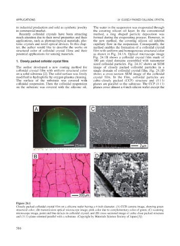
Figure 24.1
Closely packed colloidal crystal film on a silicone wafer having a 4-inch diameter. (A) CCD camera image, showing green
structural color; (B) transmission optical microscope image, pink color due to complementary color of green; (C) scanning
microscope image, point and line defects in colloidal crystal; and (D) cross sectional image of cubic close packed structure
and (111) plane oriented parallel with a substrate. (Copyright by Materials Science Society of Japan [3]).
516