
Page 261 - Optofluidics Fundamentals, Devices, and Applications
P. 261
Bio-Inspir ed Fluidic Lenses for Imaging and Integrated Optics 235
(b)
(c)
(a)
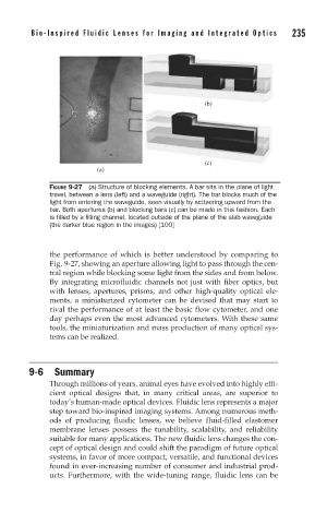
FIGURE 9-27 (a) Structure of blocking elements. A bar sits in the plane of light
travel, between a lens (left) and a waveguide (right). The bar blocks much of the
light from entering the waveguide, seen visually by scttaering upward from the
bar. Both apertures (b) and blocking bars (c) can be made in this fashion. Each
is fi lled by a fi lling channel, located outside of the plane of the slab waveguide
(the darker blue region in the images) [100]
the performance of which is better understood by comparing to
Fig. 9-27, showing an aperture allowing light to pass through the cen-
tral region while blocking some light from the sides and from below.
By integrating microfluidic channels not just with fiber optics, but
with lenses, apertures, prisms, and other high-quality optical ele-
ments, a miniaturized cytometer can be devised that may start to
rival the performance of at least the basic flow cytometer, and one
day perhaps even the most advanced cytometers. With these same
tools, the miniaturization and mass production of many optical sys-
tems can be realized.
9-6 Summary
Through millions of years, animal eyes have evolved into highly effi-
cient optical designs that, in many critical areas, are superior to
today’s human-made optical devices. Fluidic lens represents a major
step toward bio-inspired imaging systems. Among numerous meth-
ods of producing fluidic lenses, we believe fluid-filled elastomer
membrane lenses possess the tunability, scalability, and reliability
suitable for many applications. The new fluidic lens changes the con-
cept of optical design and could shift the paradigm of future optical
systems, in favor of more compact, versatile, and functional devices
found in ever-increasing number of consumer and industrial prod-
ucts. Furthermore, with the wide-tuning range, fluidic lens can be