
Page 174 - Science at the nanoscale
P. 174
10:16
RPS: PSP0007 - Science-at-Nanoscale
June 5, 2009
Nanotools and Nanofabrication
164
Optical Microscope
SEM
Electron Beam
Light Ray
Image Plane
Image Plane
Depth of Focus
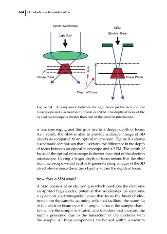
A comparison between the light beam profile in an optical
Figure 8.4.
microscope and electron beam profile in a SEM. The depth of focus of the
optical microscope is shorter than that of the electron microscope.
is less converging and this give rise to a deeper depth of focus.
As a result, the SEM is able to provide a sharper image of 3D
objects as compared to an optical microscope. Figure 8.4 shows
a schematic comparison that illustrates the difference in the depth
of focus between an optical microscope and a SEM. The depth of
focus of the optical microscope is shorter than that of the electron
microscope. Having a larger depth of focus means that the elec-
tron microscope would be able to generate sharp images of the 3D ch08
object shown since the entire object is within the depth of focus.
How does a SEM work?
A SEM consists of an electron gun which produces the electrons,
an applied high electric potential that accelerates the electrons,
a system of electromagnetic lenses that focus the beam of elec-
trons onto the sample, scanning coils that facilitate the scanning
of the electron beam over the sample surface, the sample cham-
ber where the sample is located, and detectors that measure the
signals generated due to the interaction of the electrons with
the sample. All these components are housed within a vacuum