
Page 41 - The Biochemistry of Inorganic Polyphosphates
P. 41
March 9, 2004
Char Count= 0
15:25
WU095-02
WU095/Kulaev
X-ray energy dispersive analysis 25
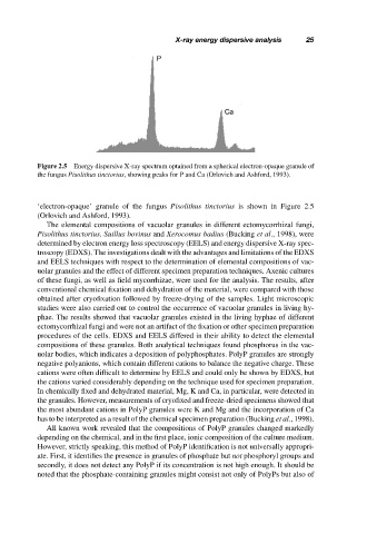
Figure 2.5 Energy dispersive X-ray spectrum optained from a spherical electron-opaque granule of
the fungus Pisolithus tinctorius, showing peaks for P and Ca (Orlovich and Ashford, 1993).
‘electron-opaque’ granule of the fungus Pisolithus tinctorius is shown in Figure 2.5
(Orlovich and Ashford, 1993).
The elemental compositions of vacuolar granules in different ectomycorrhizal fungi,
Pisolithus tinctorius, Suillus bovinus and Xerocomus badius (Bucking et al., 1998), were
determined by electron energy loss spectroscopy (EELS) and energy dispersive X-ray spec-
troscopy (EDXS). The investigations dealt with the advantages and limitations of the EDXS
and EELS techniques with respect to the determination of elemental compositions of vac-
uolar granules and the effect of different specimen preparation techniques. Axenic cultures
of these fungi, as well as field mycorrhizae, were used for the analysis. The results, after
conventional chemical fixation and dehydration of the material, were compared with those
obtained after cryofixation followed by freeze-drying of the samples. Light microscopic
studies were also carried out to control the occurrence of vacuolar granules in living hy-
phae. The results showed that vacuolar granules existed in the living hyphae of different
ectomycorrhizal fungi and were not an artifact of the fixation or other specimen preparation
procedures of the cells. EDXS and EELS differed in their ability to detect the elemental
compositions of these granules. Both analytical techniques found phosphorus in the vac-
uolar bodies, which indicates a deposition of polyphosphates. PolyP granules are strongly
negative polyanions, which contain different cations to balance the negative charge. These
cations were often difficult to determine by EELS and could only be shown by EDXS, but
the cations varied considerably depending on the technique used for specimen preparation.
In chemically fixed and dehydrated material, Mg, K and Ca, in particular, were detected in
the granules. However, measurements of cryofixed and freeze-dried specimens showed that
the most abundant cations in PolyP granules were K and Mg and the incorporation of Ca
has to be interpreted as a result of the chemical specimen preparation (Bucking et al., 1998).
All known work revealed that the compositions of PolyP granules changed markedly
depending on the chemical, and in the first place, ionic composition of the culture medium.
However, strictly speaking, this method of PolyP identification is not universally appropri-
ate. First, it identifies the presence in granules of phosphate but not phosphoryl groups and
secondly, it does not detect any PolyP if its concentration is not high enough. It should be
noted that the phosphate-containing granules might consist not only of PolyPs but also of