
Page 42 - The Biochemistry of Inorganic Polyphosphates
P. 42
WU095/Kulaev
WU095-02
Methods of polyphosphate assay
26 March 9, 2004 15:25 Char Count= 0
other phosphorus compounds. For example, the sulfate-reducing bacterium Desulfovibrio
gigas forms electron-dense granules in the cells. Energy dispersive X-ray analysis of the
granules in the cells showed that they contain large amounts of P, Mg and K. Gel elec-
trophoresis, 31 P nuclear magnetic resonance (NMR) spectroscopy and chromatographic
analyses of isolated granules revealed that they contained, instead of PolyPs, a novel metabo-
lite, which was identified as alpha-glucose 1,2,3,4,6-pentakis(diphosphate) (Hensgens et al.,
1996).
Therefore, the identification of PolyPs by X-ray techniques in some cases needs confir-
mation by using other physico-chemical methods.
2.6 31 P Nuclear Magnetic Resonance Spectroscopy
Nuclear magnetic resonance (NMR) spectroscopy is a well-established method in the study
of phosphorus metabolism (Glonek et al., 1971; Salhany et al., 1975; Burt et al., 1977; Ugur-
bil et al., 1978; Navon et al., 1977a,b, 1979; Ferguson et al., 1978; Ostrovsky et al., 1980;
Gadian, 1982; Sianoudis et al., 1986; Roberts, 1987; Shanks and Bailey, 1988; Chen, 1999).
In vivo 31 P NMR spectroscopy remains unique, being the least disruptive and quantitative
method (Gadian, 1982; Roberts, 1987; Chen, 1999).
The basic principle of the nuclear magnetic resonance (NMR) spectroscopic technique
involves measurement of the ratio frequency (rf) of the energy adsorbed by magnetic nuclei
(Roberts, 1987; Chen, 1999). NMR spectroscopy is a useful tool in analytical chemistry
for the detection, identification and structure elucidation of compounds. Phosphorus com-
pounds of living cells include phosphates, phosphonates and various esters of phosphates
and phosphonates. The chemical shift of 31 P atoms in these compounds can span over a
30 ppm range, thus making 31 P-NMR spectroscopy an attractive tool for examining phos-
phorus metabolites in microorganisms, plants and animal tissues. In addition, the method
has no problem of solvent suppression, since no water signal appears in the 31 P resonance
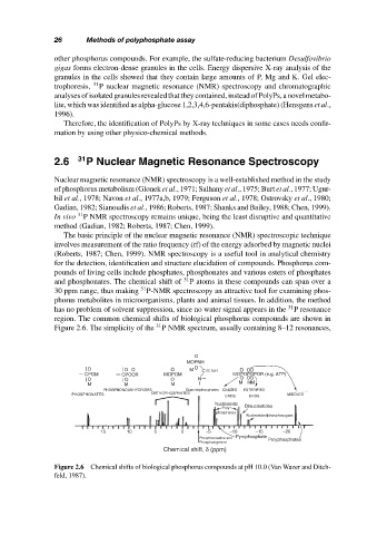
region. The common chemical shifts of biological phosphorus compounds are shown in
Figure 2.6. The simplicity of the 31 P NMR spectrum, usually containing 8–12 resonances,
O
MOPNH
O OO O M O C NH OOO
CPOM CPOCR MOPOM MOPOPOPOR (e.g. ATP)
O O O N OOO
M M M MMM
PHOSPHONOANHYDRIDES Guanidophosphates IONIZED ESTERIFIED
PHOSPHONATES ORTHOPHOSPHATES ENDS ENDS MIDDLES
Nucleoside Dinucleotides
Poly
phosphates
Nucleosidediphosphosugars
15 10 5 0 −5 −10 −15 −20
Phosphocreatine and Pyrophosphate Polyphosphates
Phosphoargenine
Chemical shift, δ (ppm)
Figure 2.6 Chemical shifts of biological phosphorus compounds at pH 10.0 (Van Wazer and Ditch-
feld, 1987).